Structure and nano nature of Bone
Bone is an extremely complex, organized and specialized connective tissue. It is highly heterogeneous, partially because of its adaptation to resist different, complex and varying stresses. The best grafts and bone substitutes are those with biomechanical and biological properties most closely resembling those of normal bone [1].
Histological Types of Bones
Bone as a tissue consists of two main types:
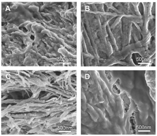
• Primary bone tissue (non-lamellar bone): This bone is also known as ‘coarse fibred’ / ‘woven’ bone or ‘immature’ bone. It is characterized by the presence of randomly oriented coarse collagen fibers.
• Secondary
bone tissue (lamellar bone): This bone is
also known as mature bone. It is characterized by
the presence of collagen fibers arranged in parallel
layers or sheets (lamellae). [2]
Bone Matrix
Bone is comprised of three basic building blocks which are nanometer in dimension - collagen fibrils, mineral plates, and a matrix of unmineralized, non-fibrillar organic material, mostly made of proteoglycans and glycoproteins. This self-assembled nanostructured ECM in bone closely surrounds and affects mesenchymal stem cell (MSC), osteoblast, osteoclast and fibroblast adhesion, proliferation and differentiation.
The bone matrix is composed of:
• Organic matter: It consists of type I collagen fibers embedded in proteoglycans and glycoproteins. The collagen fibers are made up of bundles of fibrils which act as a soft hydrogel template and resist pulling forces.
• Inorganic matter: It is made up of stiffening substances to resist bending and compression, the bone mineral is an analogue of crystals of calcium phosphate – hydroxyapatite (HA)Ca10(PO4)6(OH2). 70% of the bone matrix is composed of nanocrystalline HA which is typically 20—80nm long and 2—5nm thick. It is this association of hydroxyapatite with collagen fibers which is responsible for the hardness of bone [2].
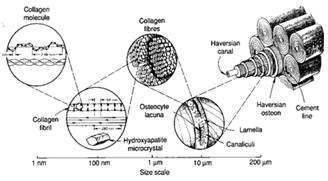
Hierarchical Structure in Bone
Bone exhibits many orders of structures: the macrostructure (cancellous and cortical bone), the microstructure (haversian systems, osteons, single trabeculae - 10 to 500 µm), the sub-microstructure (lamellae - 1 to10 µm), the nanostructure (fibrillar collagen and embedded mineral - few hundred nm to 1 µm), the subnanostructure (mineral, collagen, and non-collagenous organic proteins - below few hundred nm)
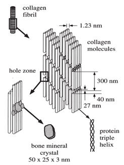
Plate-like apatite crystals of bone are positioned within the discrete spaces of collagen fibrils, limiting the possible growth of the mineral crystals, hence forcing the crystals to be discrete and discontinuous. The growth axes of the crystals are roughly parallel to the long axes of the collagen fibrils. Crystal thickness is 2–3 nm and the average lengths and widths of the plates are 50 and 25 nm respectively.
The primary organic component of the matrix is Type I collagen. Collagen molecules secreted by osteoblasts self-assemble into fibrils with a specific tertiary structure having a 67 nm periodicity and 40 nm gaps between the ends of the molecules.
Non-collagenous organic proteins, including phosphoproteins, such as osteopontin, sialoprotein, osteonectin, and osteocalcin, may function to regulate the size, orientation, and crystal habit of the mineral deposits [3].
References
[1]
H. Robert Dudley
and
David Spiro.
The fine structure of bone cells. Journal of
cell biology (1961),
vol. 11, pp.
627-649.
[2]
Aziz Nather, HJC Ong,
Zameer Aziz.Structure of bone.
www.worldscibooks.com/etextbook/5695/5695_chap01.
[3]
Jae-Young Rho, Liisa Kuhn-Spearing , Peter
Zioupos. Mechanical properties and the
hierarchical structure of bone Medical
Engineering & Physics (1998), vol. 20, pp.
92–102.