|
|
|
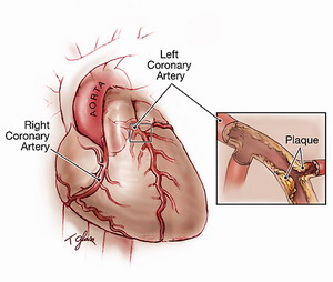
Coronary Artery Disease |
|
|
|
According to the American Heart Association, approximately 16 million people in the United States suffer from coronary artery disease (CAD), the single leading cause of death in the United States. According to present trends in the United States, half of healthy
40-year-old males will develop CAD in the future, and one in three
healthy 40-year-old women. The American Heart Association estimates that the direct and indirect cost of CAD in the United States for 2007 is more than $150 billion. |
|
|
|
|
|
Fig. 1 Coronary Artery Disease. |
|
|
|
Limitation of blood flow to the heart causes ischemia (cell starvation secondary to a lack of oxygen) of the myocardial cells. When myocardial cells die from lack of oxygen, this is called a myocardial infarction (commonly called a heart attack). It leads to heart muscle damage, heart muscle death and later scarring without heart muscle regrowth. Myocardial infarction usually results from the sudden occlusion of a coronary artery when a plaque ruptures, activating the clotting system and atheroma-clot interaction fills the lumen of the artery to the point of sudden closure. The typical narrowing of the lumen of the heart artery before sudden closure is typically 20%, according to clinical research completed in the late 1990s and using IVUS examinations within 6 months prior to a heart attack. High grade stenoses as such exceeding 75% blockage, such as detected by stress testing, were found to be responsible for only 14% of acute heart attacks the rest being due to plaque rupture/ spasm. The events leading up to plaque rupture are only partially understood. Myocardial infarction is also caused, far less commonly, by spasm of the artery wall occluding the lumen, a condition also associated with atheromatous plaque and CAD. |
 |
|
Myocardial Contrast Echocardiography |
|
|
|
Myocardial contrast
echocardiography (MCE) can detect capillary blood volume and, by
virtue of its temporal resolution, can also assess myocardial blood
flow. This imaging technique requires the delivery of a series of
high-energy ultrasound pulses to destroy contrast microbubbles in the myocardium.
Ultrasound imaging is then continued either intermittently
(during high-power imaging) or continuously (during low-power
imaging) to observe contrast intensity and microbubble velocity.
The product of peak contrast intensity and microbubble velocity gives
a measure of myocardial blood flow |
|
|
|
|
|
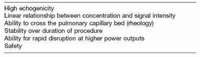
Table. 1 Desirable properties of contrast for echocardiography. |
|
|
|
Ultrasound contrast agents are gas-filled microbubbles that are administered intravenously to the systemic circulation. Microbubbles have a high degree of echogenicity, which is the ability of an object to reflect the ultrasound waves. The echogenicity difference between the gas in the microbubbles and the soft tissue surroundings of the body is immense. Thus, ultrasonic imaging using microbubble contrast agents enhances the ultrasound backscatter, or reflection of the ultrasound waves, to produce a unique sonogram with increased contrast due to the high echogenicity difference. Contrast-enhanced ultrasound can be used to image blood perfusion in organs, measure blood flow rate in the heart and other organs, and has other applications as well. A number of microbubble contrast agents are currently available in
Europe and the United States, including SonoVue (Bracco Diagnostics Inc, Princeton, NJ), Optison (GE Healthcare,
Princeton, NJ), and Luminity (Bristol-Myers Squibb Medical Imaging
Inc, North Billerica, MA; trade name Definity in United States), Imagify (Acusphere Inc, MA) |
|
|
|
|
|
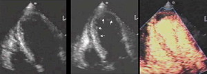
Fig. 2 Perfusion imaging in a patient with anterior ischemia. |
|
|
|
|
|
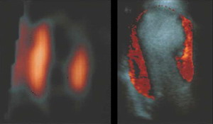
Fig. 3 Perfusion abnormality with apical myocardial infarction. |
Nuclear SPECT image (left) and color-coded myocardial contrast echocardiographic image (right). |
|
|
|
|
Imagify |
|
|
|
Imagify is an investigational new drug developed to assess perfusion using ultrasound (or echocardiography) for the detection of coronary artery disease. Myocardial perfusion is blood flow in the heart muscle and is a sensitive marker of coronary artery disease. Currently, perfusion information is not available using cardiac ultrasound but must be obtained using a nuclear stress test, which is the most frequently used imaging procedure for detecting coronary artery disease. Imagify is designed to provide real-time perfusion information with quicker results, lower cost, broader access and no exposure to radiation compared with nuclear stress tests. Imagify was designed to be used with commercially available ultrasound equipment and established imaging techniques. |
|
|

|
|
|
Fig. 4 Perflubutane. |
|
|
|