3D Geometrical Models
An
important step in modeling the arterial wall, plaque, and stent is to determine
how to mathematically describe the geometries.
In some numerical simulations, some have used a simple cylindrical model
for arterial wall. However, a more
accurate physical model can lead to more realistic computed stresses and
displacements of the arterial wall. For
instance, Kiousis et al. constructed a specific 3-D geometry of the iliac
artery based on high resolution MRI data of a patient. Then they used the non-uniform rational
B-spline (NURBS) to mathematically describe the surface. A schematic of their model is shown in Figure
4. Note that the artery is
atherosclerotic as the lumen has been substantially narrowed. The different layers of the artery is
represented by “I” for the intima, “M” for media, “A” for adventitia. The lipid pool is represented by “I-lp” [8].
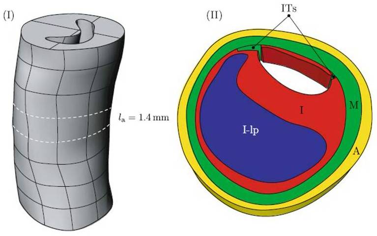
Figure 4. 3-D model of a patient’s femoral artery
constructed using high resolution MRI data [8]
A
three-dimensional model must also be generated for the stent. Using data of the structure and dimensions of
the stent acquired by a digital camera, a 3-D computer model can be constructed
by using a method called the parametrization algorithm. Essentially, this method is able to specify
the dimensions and relevant parameters of the stent geometry. This method can also help in the generation
of the mesh of the stent [8] (which will be required when using the finite
element method). Oftentimes, there is
repetition in the overall stent structure.
As a consequence, only part of the stents needs to be generated. The entire stent can then be assembled from
these parts. For instance, Figure 5 (left)
shows a meshed geometrical model of a stent cell constructed by Pro/Engineer and
imported into the commercial FEM software ANSYS [9]. Meshing is discussed in Computational
Methods.
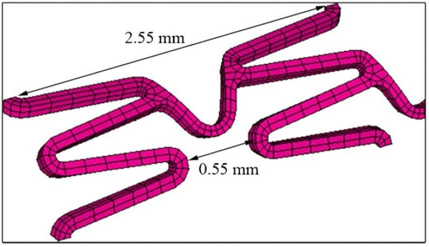
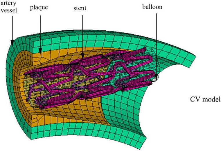
Figure 5. Meshed model of a stent cell from Wu et al (left); meshed model of stent inside a curved
atherosclerotic blood vessel (right) [9]
Figure 5 (right) shows the entire stent geometry constructed from the stent cell. The stent is situated inside a
curved blood vessel containing plaque.
The blood vessel was modeled with an inner radius of 1.5 mm, a thickness
of 0.5 mm, and a length of 12 mm. The
plaque has a length of 8 mm and a specific shape that they modeled using
uniform rational B-splines [9].
Next >>