Eye Anatomy:
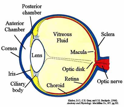
The outer shell of the eye, or globe, is primarily composed of a densely woven mesh of collagen fibers. These fibers form the sclera and cornea. The sclera is the white, optically scattering tissue composing the majority of the globe, while the cornea is the optically transparent tissue at the anterior end of the globe. This dense and stiff collagen shell provides the strength necessary to withstand and maintain pressure within the eye.
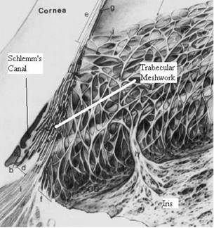
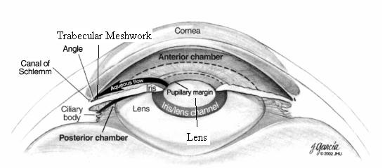
At the posterior end of the eye lies the retina and optic nerve, while the anterior portion of the eye contains the lens, iris, cornea, ciliary body, and outflow structures. The optic nerve is where damage occurs. As pressure rises within the anterior chamber, it is ubiquitously distributed through the vitreous gel which fills the rest of the eye posterior to the lens. The outflow structures are defined as the tissues the aqueous humor must pass through upon exiting the eye. They include the trabecular meshwork, juxctacanalicular tissues, inner wall of schlemm’s canal, schlemm’s can, and the collector channel which lead to the episcleral venous system of the eye.
To better understand the importance of these outflow structures with respect to femtosecond laser glaucoma treatment, it is important to understand in more depth the fluid dynamics associated with the aqueous humor.