


|
|
|
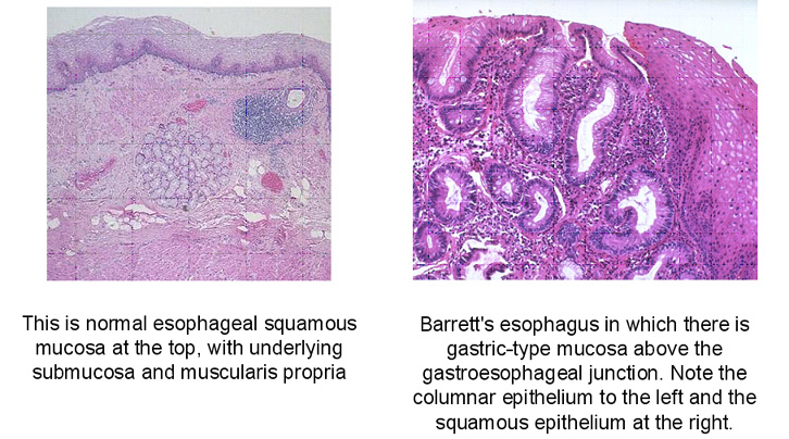
Two criteria are required for the diagnosis of
Barrett esophagus: endoscopic evidence of columnar epithelium above the
gastroesophageal junction and histologic evidence of intestinal
metaplasia in the biopsy specimens from the columnar epithelium (6).
Grossly, Barrett esophagus is recognized as a red, velvety mucosa
located between the smooth pale pink esophageal mucosa and the light
brown gastric mucosa. It may exist in tongues or patches extending from
the gastroesophaeal junction or as a broad irregular circumferential
band displacing the squamoucolumnar junction several centimeters
cephalad. Microscopically, in Barrett esophagus the esophageal squamous
epithelium is replaced by metaplastic columnar epithelium containing
intestinal goblet cells.
|
|
|
|
|
|
|
| |
|
|