


|
|
|
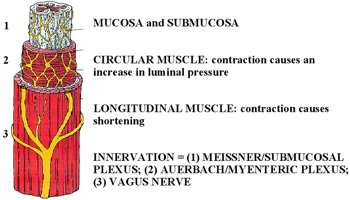
The wall of the esophagus is composed of four
layers (inner to outer): mucosa, submucosa, muscularis propria and
adventitia, reflecting the general structural organization of the entire
gastrointestinal tract. The mucosa is composed of three components
(inner to outer): a non-keratinizing stratified squamous epithelium, a
lamina propria and the muscularis muscosa. The submucosa is composed of
loose connective tissue, blood vessels, lymphatics, lymphoid follicles,
the meissner plexus of nerves and submucosal glands. The submucosal
glands secrete mucus which functions to lubricate the esophagus and
protect the epithelial layers from the harsh environment to which it is
exposed, e.g., food and gastric acid. The muscularis propria consists
of an inner circular muscle layer and an outer longitudinal muscle layer
with an intervening myenteric (auerbach) plexus. Contraction of the
circular muscle layer causes an increase in luminal pressure, while
contraction of the longitudinal muscle layer causes shortening of the
esophagus. The muscularis propria of the esophagus is unique in that
the proximal 1/3 is composed of skeletal muscle, the middle 1/3 is
composed of both smooth and skeletal muscle and the distal 1/3 is
composed of only smooth muscle. The adventitia is the connective tissue
fascia layer that surrounds the esophagus (1). |
|
|
|
|
|
|
| |
|
|