
The Method
Seven patients (3 males and 4 females) were used in the study with an average age of 11 (4-19 years old). 3 Biopsies were performed 7-8 prior to the actual cystoplasty. This would enable the a small sample of autologous cells to be collected. A 1-2 cm 2 sample was obtained. The cells were cultured and placed on various scaffolds. Two types of scaffolds were used ranging from 70 to 150 cm 2, as mentioned previously; a collagen-PGA scaffold was employed along with a template that was custom taylored to each patient's bladder size. Using CT scans, 3D images were obtained along with thicknesses required for the tissue. As shown in figure 4, each of the constructed bladders were customized according to the patient's age and pelvic cavity.3
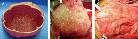
Figure 4: The engineered bladder. (A) Scaffold with cells; (B) Engineered bladder connected to bladder with sutures; (C) Implant covered with fibrin glue and omentum (3)
In figure 4A, we see the engineered bladder consisting of both smooth muscle cells (outer layer) and urothelial cells (inner layer). Over 70 plates for each cell type was used with each plate containing an average of 10 million cells.3 The scaffold was coated with these cells at appropriate times and allowed to attach to them. The scaffolds were then incubated for 3-4 days until it was ready for implantation.3