 |
|
|
Home | Clinical Background and Need | Engineering Principles and Clinical Considerations | Commercial Devices | Upcoming Advancements |
| Clinical
Background and Need
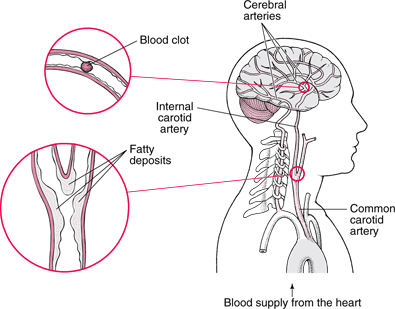
Definitions A stroke is defined as the acute loss of blood perfusion to the brain. Strokes are classified as ischemic or hemorrhagic(1). Hemorrhagic strokes involve intracerebral or subarachnoid bleeding - bleeding within the brain or bleeding in the subarachnoid space, respectively (2). Hemorrhagic strokes have different causes and treatments than ischemic strokes and are out of the scope of this discussion. For basic information on hemorrhagic strokes, visit Merck's Article on Hemorrhagic Stroke. Ischemic strokes involve occluded blood arteries that route blood into the brain and are the focus of treatment for this webpage. More technically, ischemic stroke is defined as focal brain infarction that causes neurological deficits persisting longer than one hour (3). Causes of Ischemic Stroke Thrombosis: Thrombosis is essentially a blood clot. Thrombosis forms for various reasons, most commonly due to slowed blood flow due to atherosclerosis or ruptured atheromas (see atherosclerosis below). Slowing of blood flow decreases the shear stresses seen by platelets and red blood cells, which in turn makes clotting easier to achieve. The thrombus forms in an artery supplying blood to the brain and restricts blood flow at that location. In the image to the right, "Blood clot" is an example of a thrombosis (if it originated in that particular location in the cerebral artery). Embolism: Thrombus or pieces of atheromas or plaques can originate upstream, usually in the heart, on a heart valve (especially prosthetic valves), or aorta, and move downstream and lodge in arteries supplying the brain. In the image to the right, if "Blood clot" had originated upstream from its location, such as in the heart, then it would be classified as an embolism. Lacunar infarct: Lacunar infarcts are small (less than 1.5 cm) infarcts resulting from nonatherothrombotic obstruction of small arteries deep in the brain. The usual cause is the degeneration of the media of the small arteries- thrombus is not the cause. Atherosclerosis: Atheromas or plaques themselves may grow large enough to occlude arteries, leading to ischemic stroke. In addition (and more relevant to the focus of this webpage), these regions are likely to form thrombus because of the slowed blood flow due to the constriction of the vessel. "Fatty deposits" in the image to the right is an example of atheromas (3), (4). Image from (4). Clinical Treatment of Ischemic Stroke Anticoagulants/Antiplatelet Drugs: Most patients are immediately treated with antiplatelet drugs like aspirin. Antiplatelet drugs prevent platelets from aggregating and thus minimize clotting (or deteriorate existing thrombus). If symptoms persist or worsen, patients are often given anticoagulant drugs like heparin. Anticoagulants prevent clotting factors and thus also minimize clotting (or deteriorate existing thrombus). Thrombolytic drugs and anticoagulant or antiplatelet drugs are not used simultaneously due to the greatly increased risk of hemorrhaging in the brain (4). Thrombolytic Drugs: Thrombolytic drugs are drugs that break up clots (thrombosis) and thus restore blood flow to the brain. This therapy is well-proven via 21 clinical trials enrolling 7,152 patients. Various drugs, doses, time windows, and modes of administration were investigated in these trials. The following results have been accepted from these trials: intravenous administration of thrombolytic drugs within the first 3 hours of ischemic stroke onset offers benefits for nearly all patients. Intravenous administration within 3-4.5 hours of ischemic stroke onset offers moderate benefits to nearly all patients. Intra-arterial administration with 3-6 hours of ischemic stroke onset offers moderate benefits to nearly all patients with large cerebral artery thrombotic occlusions (5). The most commonly used drug is tPA, tissue plasminogen activator or rt-PA, recombinant tissue plasminogen activator. tPA and rt-PA can cause bleeding in the brain (as well as elsewhere in the body) and have many contraindications, such as, intracranial hemorrhage on CT scan or history of intracranial hemorrhage, multilobar infarct, platelet count, use of heparin within 48 hours, and others. Therefore, they are not used in most patients (3), (4). In addition, recanalization rates achieved with intravenous rt-PA for proximal, large arterial vessel occlusion are poor: 10% for internal carotid artery occlusion and 30% for middle cerebral artery occlusion. Intravenous thrombolysis is more effective in smaller, more distal vessels. Thrombolytic drug injections are ineffective in dissolving platelet-rich clots and have lengthy times to recanalization. Additionally, reocclusion at the location of obstruction occurs in 34% of patients injected intravenously and 17% of patients injected intra-arterially (6).Mechanical Thrombectomy: Mechanical thrombectomy is the endovascular, mechanical disruption and retrieval of thrombosis at the site of occlusion. This therapy yields much higher recanalization rates in patients with thrombosis in larger, more proximal arteries. There are currently two mechanical thrombectomy devices approved by the FDA for use in the United States. These devices are approved for patients with stroke symptoms that have not responded to thrombolytic drug therapy or are contraindicated for such therapy. Recanalization rates in these devices range from 50% to 80% (6). Clinical Need Approximately 795,000 (625,000 ischemic) strokes occur in the United States every year. 15 million strokes occur worldwide every year as estimated by the World Health Organization (1). Less than 5% are being treated with drug therapies (7). 1) Jauch E C, Kissela B. Acute Stroke Management. emedicine from WebMD. 2009. 2) Giraldo E A. Hemorrhagic Stroke. Merck Online Manual. 2007. 3) Giraldo E A. Ischemic Stroke. Merck Online Manual. 2007. 4) Giraldo E A. Ischemic Stroke. Merck Online Manual. 2007. [Different than (3)] 5) Saver J L, Kalafut M. Thrombolytic Therapy in Stroke. emedicine from WebMD. 2010. 7) The Penumbra Pivotal Stroke Trial Investigators. The Penumbra Pivotal Stroke Trial. Safety and Effectiveness of a New Generation of Mechanical Devices for Clot Removal in Intracranial Large Vessel Occlusive Disease. Stroke, 40: 2761-2768. 2009. |