Melanoma is a form of skin cancer that is often defined in medical textbooks as being a "malignant tumor of melanocytes," and while it only encompasses about 5% of all skin-cancer cases, it is also the most deadly, accounting for almost 80% of all skin-cancer related deaths around the world. Although 5% may not seem like a large percentage, it must be kept in mind that skin cancer is by far the most common form of cancer. According to the Skin Cancer Foundation, more than 3.5 million cases in two million people are diagnosed annually with skin cancer, and of that population, about 68,720 will be diagnosed with melanoma, with nearly 8,650 resulting in death.
The high mortality rate of melanoma is directly related to the lack of appropriate patient screening and early detection techniques. Effective prognosis of melanoma is typically related with early diagnosis, but at the moment, there is no substantial method that can truly be deemed precise. As of now, many diagnoses are done by visually inspecting lesions, and conducting biopsies when a suspicious abnormality is found. Unfortunately, even the most expert clinicians with many years of experience can have difficulty in differentiating cutaneous melanoma from a benign lesion. This makes the technique very prone to false-postives, or even worst, false-negatives.
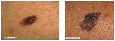
Figure 1: Visual comparison of a benign lesion, dysplastic nevus, (left) and a malignant, lentigo maligna, melanoma (right). Both look very similar and can often be confused for one another.
Depending on the experience and skill of the clinician, it can take from 5 to even 100 benign lesion biopsies in order to find a single melanoma. This in turn creates a very excruciating environment for patients, who after having so many tissue samples taken from their body, start feeling like "a pile of swiss cheese." Given the time-sensitive nature of melanoma diagnostics and prognosis, it is clear that current methods are not very ideal for solving the problem at hand.
Because of the demand for a quick and efficient technique to diagnose melanoma, there have been much research put into finding a good alternative, the most promising of which stem from optical biomedical engineering techniques.
Optical diagnostics of living tissues is largely based on the principles behind spectroscopy. Spectroscopy is the study of molecular structure and dynamics via the interaction of light with matter. The wavelength (energy) of the light determines how it interacts with the matter via absorption, emission, and scattering. These interactions can be then used as a probe in order to obtain chemical information about the sample such as the identification of constituents or the concentration of constituents.
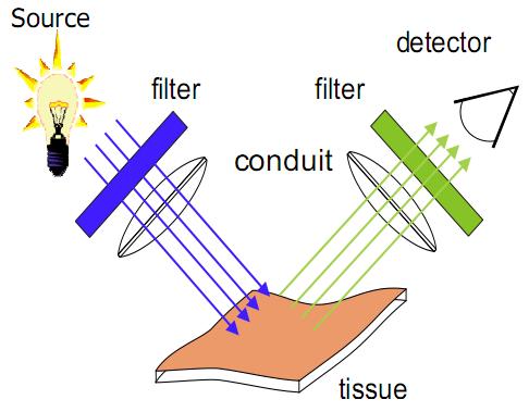
Figure 2: Basic engineering design behind tissue spectroscopy.
Figure 2 gives an illustration of the basic engineering design behind tissue spectroscopy. In its most simple form, a light source with a known wavelength and intensity is applied to the tissue surface. Depending on the wavelength and target chromophore, a series of photon-tissue based interactions will occur during which some light photons will be scattered, while others will be absorbed. The amount that is reflected back to the detector can then be used to determine qualitative and quantitative properties of the tissue in question.
Optical spectroscopy has been used in many scientific fields in order to interrogate unknown substances without physically perturbing them. Optical diagnosis of melanoma, in essence, is just another another practical application of optical spectroscopy where the goal is to identify suspicious tissue compositions before life-threatening metastasis occurs. Its advantages lie in its ability to efficiently and effectively quantify and qualify tissue data through non-invasive means.
The chromophores of interest when measuring optical diagnostic data are:
From these information, a detailed measure of the hemodynamic, energetic, and morphological states of a tissue sample can be made.
There are many forms of optical diagnostic techniques for detecting melanoma, and on this website we will cover three very prominent implementations of this technique: Diffuse Optical Spectroscopic Imaging (DOSI), Spatial Frequency Domain Imaging (SFDI), and Laser Speckle Imaging (LSI).
It should noted that all three technologies are currently under development at the Beckman Laser Institute (BLI) in Irvine, California. For more in-depth and up-to-date news and journal articles, please visit their website at http://www.bli.uci.edu/.
John Nguyen 2010
BME 295