Diffuse Optical Spectroscopic Imaging (DOSI) is a non-invasive optical diagnostic technique that can quantify the absorption and scattering coefficients of tissues up to several centimeters deep. By measuring these optical properties, quantifiable and qualitative information about the target tissue can be ascertained. The beauty of DOSI is that it is fundamentally based on basic spectroscopic theories. To be more specific, DOSI is based on optical spectroscopy in the approximate 800nm to 2500nm near-infrared (NIR) region of the electromagnetic spectrum. It works by interrogating the target tissue with NIR light, detecting the remitted photons, and analyzing the remission via a set of mathematical photon transport models based on Beer-Lambert Law modified to predict multiple photon scattering and diffusion in living tissues. From this, various useful information such as tissue oximetry can be used to diagnose conditions such as melanoma.
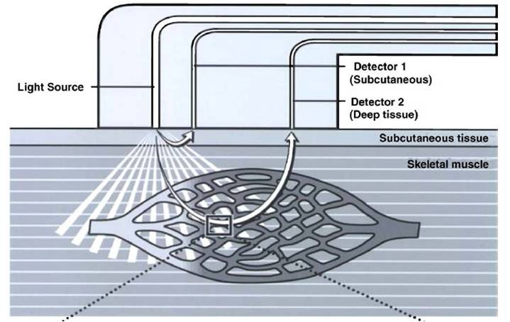
Figure 1: Basic engineering design behind DOSI.
As can be seen in Figure 1, a typical DOSI instrument consists of a tunable NIR laser light source and various photon detectors placed at differing distances away from the source. When laser light enters the region of interest, it begins to scatter and diffuse in a "banana" shaped fashion throughout the tissue. This "banana" shaped light diffusion path is the reason why detectors closest to the source are able to analyze superficial portions of the tissue, while the furthest detectors are capable of a deeper interrogation. Of course there is a limit to how far away the detectors can be. If they are too far away, all of the photons from the light source would be diffused and would be undetectable by the probe. Because of this limit, DOSI systems tend to have a very restricted field of view compared to other optical diagnostic techniques. The plus side to this is that DOSI instrumentation also tend to be relatively small and portable.
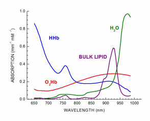
Figure 2: Absorption spectra of various chromophores at different wavelengths including NIR.
As was previously stated, DOSI is based on optical spectroscopy which assumes that every chromophore in the target tissue absorbs the most light at their own unique and specific wavelength. This can be seen in Figure 2. For example, water absorption of NIR light is highest around the 980nm range. Another thing to notice is that oxygenated and de-oxygenated hemoglobin have dramatically different absorption profiles, and are thus relatively easy to differentiate from one another. By measuring the tissue-photon interaction during DOSI studies, chromophore content and composition can be individually separated and analyzed to determine differences between benign lesions and malignant melanoma.
John Nguyen 2010
BME 295