Laser Speckle Imaging (LSI) is a non-invasive blood flow imaging technique that can provide information on the state of biological tissues and the efficiency of disease treatment. Unlike DOSI and SFDI which are aimed at diagnosing melanoma, the goal of LSI is more oriented towards evaluating the process of non-invasive melanoma treatments such as Photodynamic Therapy (PDT). Also based on the absorption and scattering foundation behind many optical diagnostic techniques, LSI focuses on interpreting the speckle pattern phenomenon that occurs when electromagnetic light waves interfere with one another to produce optically visible effects in both the spatial and temporal regions of the remitted reflectance pattern. Analysis of these speckle patterns can result in quantitative wide-field imaging data pertaining to blood flow velocity mapping. This in turn is very useful for evaluating the efficiency and extent of the melanoma treatment.
The basic engineering design of an LSI system is very similar to that of SFDI, but with a 30mW, 63nm helium-neon laser used in place of the digital projector. The widefield imaging aspect is produced by rendering the laser divergent through of a series of lenses. Images are captured by a commercial grade CCD camera and data is processed via an onboard computer system.
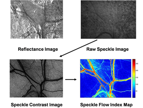
Figure 1: Example data flow for LSI.
Figure 1 outlines the basic data flow for an LSI system. A CCD camera captures the reflectance of a standard lamp illumination. Laser excitation is introduced to create the raw speckle image that can be visibly described as "grainy". Using a computer to process the raw data with a mathematical convolution filter, a speckle contrast image can be obtained, which is then used to create a speckle flow index map that describes the blood flow velocity in the region of interest.
John Nguyen 2010
BME 295