The therapeutic product is a hSLo cDNA gene that encodes the alpha subunit of the smooth muscle cell potassium ion channel known as the Maxi-K channel.
The cDNA fragment is inserted into a plasmid construct which is commercially available (pVAX by Invitrogen). The plasmid construct (Figure 2) contains both the gene encoding Maxi-K channel as well as promoter sequences needed for transcription. A phosphate buffered saline solution containing the plasmid construct is then administered into the corpus cavernosum.
The advantage of this plasmid construct is that no viral vector is used to transfer the gene and thus avoids the adverse immune responses and inflammation typically caused by viral vectors.
The disadvantage is that the efficiency of the transfection (proportion of recombinant cells in the host) is low compared to viral vectors.
Proposed mechanism of the therapeutic product
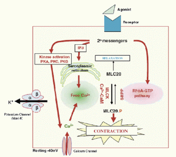
(Figure 3a) shows the regulatory mechanism for smooth muscle cells relaxation in the corpora cavernosa. The K+ channels play an important role in regulating the contraction and relaxation of the smooth muscle cells in the corpora cavernosa of the penis. Their primary effect is to modulate the influx of calcium ions into the cells which in turn regulate the degree of smooth muscle contraction.
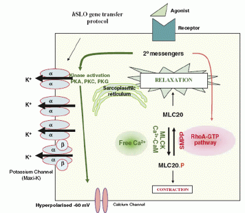
Increased Maxi-K channel activity(Figure 3b) is associated with corporal smooth muscle cell relaxation and penile erection.The ion channels signals are propagated via gap junctions in the penile corporal smooth muscle cells network. Thus increased concentration in K channel activity in one site can be propagated via these gap junctions, allowing the corporal tissue to functions as a whole unit even after severe nerve loss.
This is also why unlike cancer cell gene therapy for instance, a low rate of recombinant cell transfection is not necessary when using the plasmid mediated gene transfer.
In conclusion, the use of ion channel therapy via upregulation of the genes encoding the hsLo subunit is based on the principle of allowing the rapid and robust relaxation of the corporal smooth muscle cells.
 |
| Top Figure 3a: Left figure shows relationship of the smooth muscle cell to the potassium ion channels, intracellular
calcium ion concentrations,secondary messengers and other proteins. Smooth muscle contractility is regulated primarily by intracellular Ca ions, the temporal control of calcium ion entry by potassium channel activity regulates the tone of the smooth muscle cell. |
 |
| Bottom Figure 3b: Increased K channel activity via insertion of Maxi-K channels into smooth muscle cell. Three additional Maxi-K channels are shown in the cell membrane. In the presence of
an appropriate neural or ionic signal, the potassium channels open, hyperpolarize the cell to approximately -60 mV, and
inhibit the influx of calcium ion, thus causing the cell to relax. |
|
