Stem Cell Culturing:
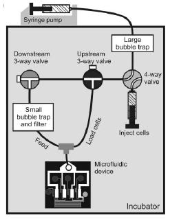
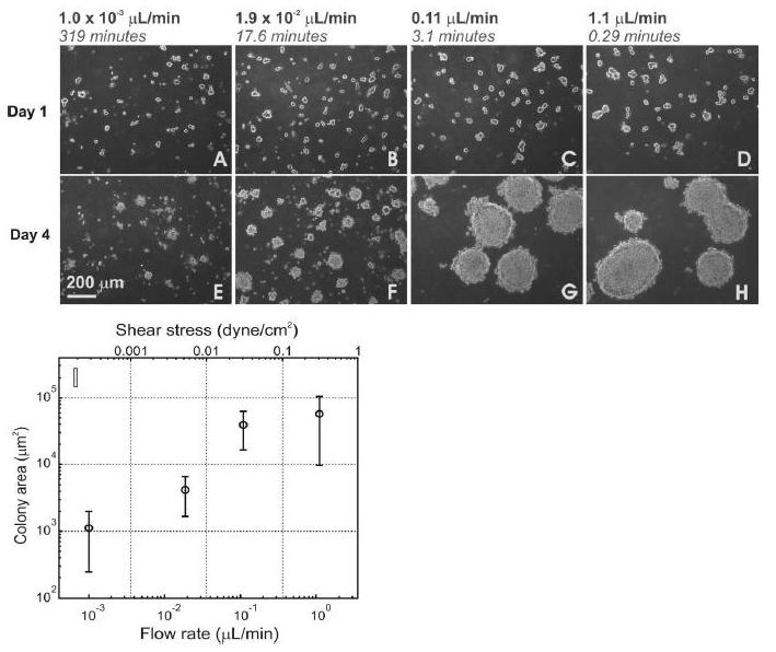
The work conducted by Dr. Beebe’s lab at the University of Wisconsin-Madison, paved the way for implementing microfluidic devices for the use with stem cells. His group published the first successful culturing of embryonic stem cells on both a mouse feeder layer and a layer of matrigel, which is a gel derived from mouse sarcomas. The advantage for being able to culture stem cells in microfluidic devices is that the controlled fluid dynamics within the channels of the device allow for microenvironments to be held stable which is very similar to that in developing embryos. However, another approach has been taken by the Voldman lab at MIT. The work shown by their group indicates that a stem cell culture can be grown more successfully in a perfusing environment. The microfluidic device which they developed enables varying flow rates to be administered by a syringe pump as well as the generation of a logarithmic gradient profile of soluble molecules. The experiments showed that increased flow rates yielded higher proliferation of colonies. Therefore a positive relationship exists between the shear forces exerted on the cells and their ability to thrive in culture. Although this relation will plateau and certainly decrease with much higher flow rates, being able to guide cells behavior by external forces is very beneficial. The importance of these advancements is that the quantification of culturing conditions sets a basis for optimization to occur for specific cell types. Also, the ability to expand the device to yield high-throughput screening significantly enables stem cells to become a more cost-efficient therapy.
Guiding Stem Cell Differentiation:
Stem cells are highly sensitive to their microenvironment which includes both their substrate and the chemicals they come in contact with. Varying the substrate which a cell colony grows on can have a tremendous effect on its phenotypic response. These responses can be monitored and characterized by analyzing the reaction of stem cell differentiation on different substrates. This work was done by Dr. Bhatia’s lab at UC San Diego. They utilized a microarray of extra-cellular matrix and were able to determine optimum coating conditions. The high-throughput ability of this device enables for very efficient screening and optimizing of stem cell substrate conditions. The developed microarray uses 1000 times less protein than conventional methods while allowing 32 different combinations of proteins to be mixed. 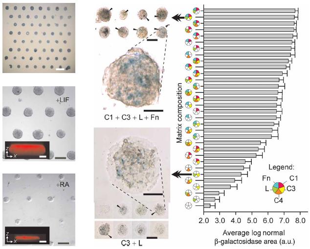
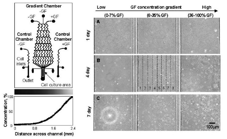
Stem cell differentiation is also heavily controlled by the soluble factors in the surrounding media. These factors can be uniformly dispersed or exist in a gradient at any range of concentrations. Microfluidics provides a perfect platform to generate and maintain stable gradients to study and optimize conditions for stem cell differentiation. Dr. Jeon’s Group at UC Irvine utilized a microfluidic gradient generator, depicted below, to study the differentiation of neural stem cells in a gradient of soluble growth factors. It was seen that within a device, areas with low growth factor concentration yielded high percentage of differentiation into astrocytes while areas with high concentration of growth factors resulted in low differentiation but high proliferation. These findings serve as a great basis where within a culture, stem cells can be differentiated and used for a therapy as well as replenish itself to serve as a constant source.