Ultrafast Laser Basics
The term ultrafast lasers applies to lasers which, rather than emitting light in form of a continuous wave (cw), emit a series or train of pulses, each one of which lasts less than a nanosecond. The following section provides a brief summary of how ultrashort pulses are generated, as well as a short overview of current ultrafast systems.
Generating Laser Light

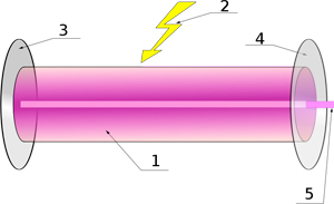
Figure 1. Schematic drawing of a laser cavity. 1. Gain medium, 2. Laser pumping energy, 3. Highly reflective mirror, 4. Partially transmissive mirror (Output coupler), 5. Laser beam
At its most basic level, a laser consists of a gain medium inside an optical cavity with a highly reflective mirror on one end, and a partially transparent mirror on the other end. The gain medium can be a gas, a mixture of gases, or a solid-state material such as a crystal or a semiconductor. Atoms inside the gain medium are excited, or pumped, by means of an external energy supply. This can be achieved through various means depending on the medium and the type of laser, ranging from applying an electrical current to using a flash lamp or even another laser. The pumping process causes the outer electrons of the gain medium's atoms to jump to a higher, excited, energy state. Because the excited state is unstable, the electrons return to their ground state after a short while, releasing excess energy in the form of a photon. When this photon strikes another excited electron, it causes the electron to return to its ground state immediately, releasing a second photon of the exact same phase and wavelength as the incoming photon. This process occurs many times in the cavity, creating a photon avalanche, which eventually gets reflected by the highly reflective mirror at the end of the cavity.
On its way back through the cavity, more photons are released, until the photons reach the partially transparent mirror at the other end. This mirror typically has a transmissivity of between 1 to 2 %, permitting a small amount of photons to leave the cavity in the form of the characteristic monochromatic, coherent beam of light.
The wavelength of the laser beam is determined by the type of gain medium, as the individual photon wavelength depends solely on the amount of energy emitted when returning to the ground state as determined by the de Broglie relation1 E = hf, where E is the energy, f is the frequency and h is Planck's constant.
Prominent examples of such basic cw lasers include gas lasers such as Helium-Neon (He:Ne) and Argon Ion lasers. Gas lasers can typically emit more than one frequency, referred to as a mode.
Generating Ultrashort Pulses
Creating pulses can be achieved either actively or passively. If long pulses are required, a mechanical shutter such as a rotating cogwheel can be used to chop the continuous beam into pulses. This is insufficient for shorter pulses however, mainly due to the limited angular velocity of any mechanical wheel. Non-mechanical switches, such as Pockels cells and acousto-optic modulators can be used to create shorter pulses, but in order to achieve sub-nanosecond pulses passive pulse creation is required. To passively create short pulses, lasers exploit an effect called Kerr-Lens modelocking (KLM) which allows the creation of pulses in the picosecond, femtosecond and recently even the attosecond range2.
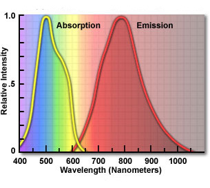
To illustrate the principle of modelocking, consider the Titanium-Sapphire (Ti:Sapphire) laser, which is one of the most commonly used short-pulsed lasers. It features a very broad emission spectrum ranging from 600 nm into the infrared range up to about 1050 nm. This spectrum, together with its broad absorption spectrum which allows for a multitude of pumping lasers to be used with this crystal makes the Ti:Sapphire crystal ideally suited for the creation of femtosecond pulses.

Figure 1. Absorption and emission spectra for a Ti:Sapphire laser. The broad absorption spectrum allows for a wide range of pumping lasers, while the wide emission spectra provides a large amount of modes that can be combined to achieve femtosecond pulses.
In order to create modelocked pulses, the various longitudinal modes of the laser cavity are locked, creating a fixed relationship between their individual phases. If the length of the laser cavity is L, the frequency difference between two adjacent modes is c/2L.
Locking phases creates a constructive interference pattern between the various modes, resulting in a pulsed light emission. The final pulse length is determined by the amount of modes that are locked together as t=0.44*2L/c 3.
The pulse width as a function of the amount of modes will therefore decrease if more modes are locked. Taking the frequency separation of two modes into account, the bandwidth required for the creation of N modes is given by N*c/2L. The maximum amount of modes that a laser can support is therefore determined by its bandwidth. The Ti:Sapphire laser with its broad emission range of over 400 nm can theoretically achieve pulse lengths in the single-digit femtosecond range.
Modelocking is achieved by means of a nonlinear optical effect called Kerr-effect. The refractive index of the Ti:Sapphire crystal is a function of the light intensity and increases at higher light intensities. Assuming a gaussian beam profile, the highest intensities can be found in the center of the beam. Due to the higher index of refraction resulting from the Kerr effect, the center of the beam travels more slowly than the periphery of the beam, effectively creating a lensing effect. Because the intensities of the continuous-wave (cw) mode are lower than the peak intensities of a pulsed beam, the cw beam undergoes less focusing. Using a slit to block the outer regions of the beam the cw mode is suppressed, resulting in a self-amplifying pulsed beam4. This technique, called Kerr-lens modelocking, does not require moving parts such as the rotating cogwheel or active components that have to be electronically controlled like the AOM. KLM is therefore sometimes referred to as "self mode locking".
Using KLM the Ti:Sapphire laser is theoretically capable of reaching its minimum pulse length. The actual pulse length that can be achieved in a real laser however depends on other factors, such as the group velocity dispersion, which is discussed in more detail in the last section.
Laser-Tissue Interaction

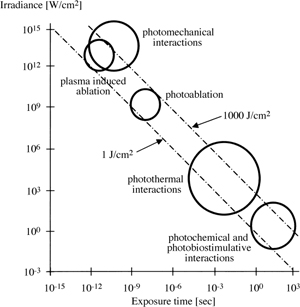
Figure 3. Laser-Tissue interactions as a function of pulse duration and laser power. The two lines indicate the 1 and 1000 J/cm2 irradiance limits.
When laser light strikes tissue, various interactions occur depending on the intensity of the light, ranging from simple thermal interaction due to photon absorption to non-linear effects such as Second Harmonic Generation (SHG) and Laser-Induced Optical Breakdown (LIOB). Additionally, several other non-destructive interactions may occur, none of which are of interest for surgical applications. The relevant interactions are limited to only a handful of different effects, illustrated in Fig. 3. Ultrashort laser systems exploit photodisruption and plasma-mediated ablation, while surgical cw lasers such as the Excimer UV laser used for LASIK surgery relies on lower-tier interactions like photoablation.
Laser-Induced Optical Breakdown
Two destructive effects occur primarily during LIOB: Plasma-mediated ablation and photodisruption. LIOB requires extremely strong electrical fields in and above the megawatt range, which are normally associated with extremely high powered cw lasers. On account of their raw power however, these lasers are completely unsuitable for surgery, as they deposit massive amounts of energy into the target volume, causing excessive heating and vaporization of large volumes of tissue. Because power is defined as energy over time however, ultrashort pulse durations allow for extremely large-scale fields while depositing only microjoules of energy. Even though the field strength is high enough to allow for LIOB, the total amount of energy deposited is small enough that there is very little interaction outside the focal volume. It is this effect that makes ultrashort pulses well suited for high-precision microsurgery.

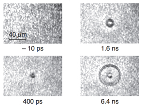
Figure 4. Shockwave expansion in water following a 100fs pulse at 800nm as imaged by Schaffer et al 5.
Consider a femtosecond-range pulse interacting with a piece of tissue. In the focal volume of the beam, the electric field strength increases rapidly and by many orders of magnitude, exceeding the strength of the field that binds the electrons to their host nuclei. As a result, electrons are stripped from their host atoms, forming free electrons that are further accelerated by the laser-induced electrical field. This results in an electron avalanche and the subsequent ionization of atoms in the focal volume, leading to the formation of a plasma. This plasma has extremely high absorption rates and will absorb most of the energy from the laser beam. As a result, the temperature of the plasma will rise rapidly, causing the newly formed plasma bubble to expand at high speeds. Tissue is now being turned into plasma in a process called plasma-mediated ablation. The expansion generates supersonic shockwaves that ripple through the tissue, causing mechanical damage. This process is known as photodisruption. Secondary effects such as plasma bubble oscillation and the formation of plasma jets as the bubble collapses may cause further damage.
Because LIOB requires extremely high field strengths, it only occurs in the very focal volume of the laser beam, allowing for highly precise ablation and disruption in the micron range. Furthermore, because thermal interaction requires mechanical coupling between vibrating atoms or molecules, there is no direct thermal coupling between the beam and the tissue on account of the pulse duration being faster than the speed of thermal vibrations.
Commonly used systems today include lasers using various elements doped with Neodymium (Nd), such as Nd:YAG, Nd:Glass or Nd:YLF. Emission and absorption spectra, stability, resistance to destructive effects inside the lasing cavity, laser-tissue interaction at the desired wavelength and of course cost are some of the factors used to determine the optimal lasing medium for a specific surgical task.