 |
Background
What are urinary tract calculi?
Chemical ions, molecules, and compounds normally exist in urine (i.e., calcium, oxalate, phosphate, uric acid). When the concentrations of these substances are high, they combine to form crystals. Under appropriate environmental conditions and with the aid of debris in the urinary tract, these crystals will grow and aggregate together resulting in the formation of larger crystals and eventually stones.
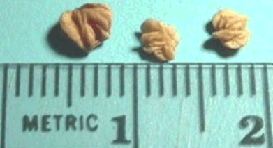
Why should I be worried of stones? How can I know I have them?
If a stone grows large enough it can get caught in the kidney or the ureter (the tube that drains the kidney into the bladder). Once it gets caught, the stone may partially or completely block the flow of urine. This blockage causes pain that is usually felt in the middle of the back or side and may radiate toward the groin. Sometimes the pain can be so severe as to cause nausea and vomiting. Fevers and chills may accompany a stone that is associated with infection. If a stone that is blocking urine flow is left untreated it can cause damage to the kidney or ureter.
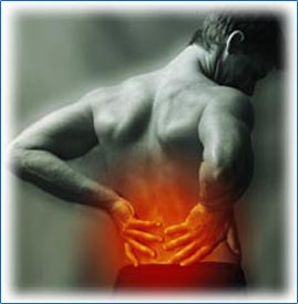
Some stones may not cause any symptoms at all. Stones in the bladder may cause pain in the lower abdomen. Stones that obstruct the ureter or renal pelvis or any of the kidney's drainage tubes may cause back pain or renal colic. Renal colic is characterized by an excruciating intermittent pain, usually in the flank (the area between the ribs and hip), that spreads across the abdomen, often to the genital area and inner thigh. The pain tends to come in waves, gradually increasing to a peak intensity, then fading, over about 20 to 60 minutes. The pain may radiate down the abdomen toward the groin or testicle or vulva.
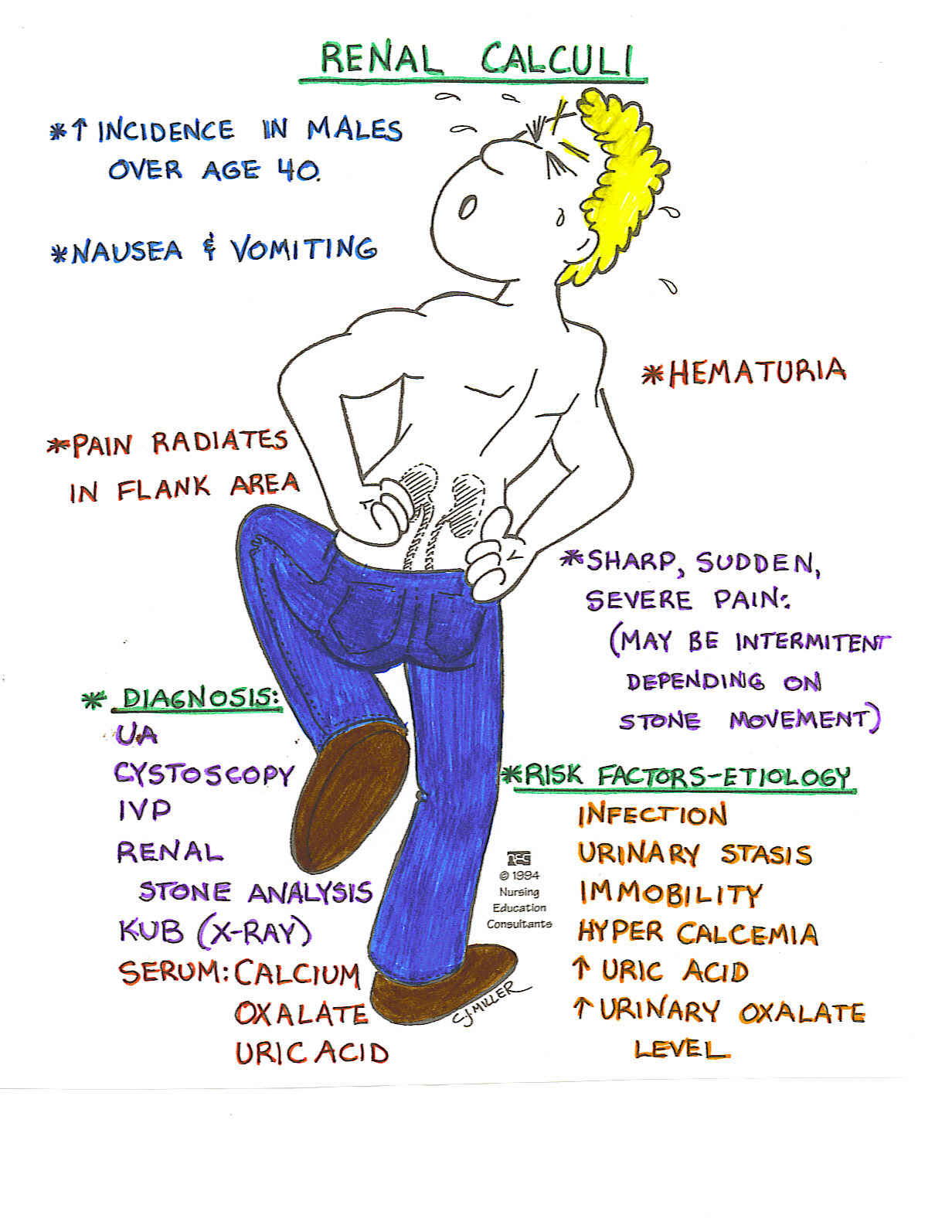
Other symptoms include nausea and vomiting, restlessness, sweating, and blood in the urine. A person may have an urge to urinate frequently, particularly as a stone passes down the ureter. Chills, fever, and abdominal distention sometimes occur.
How are stones diagnosed?
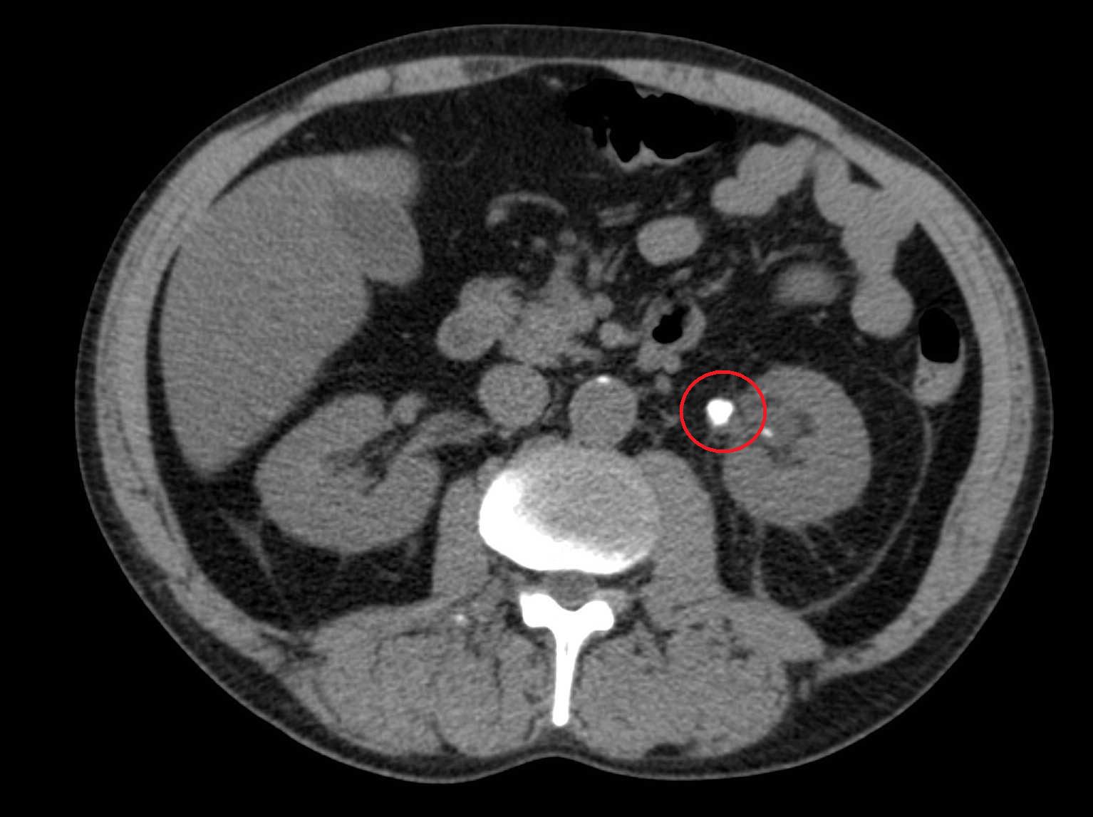
Helical (also called spiral) computed tomography (CT) that is done without the use of radiopaque contrast material is usually the best diagnostic procedure. CT can locate a stone and also indicate the degree to which the stone is blocking the urinary tract. CT can also detect many other disorders that can cause pain similar to the pain caused by stones. The disadvantage of CT is that it exposes people to radiation. Still, this risk seems prudent when possible causes include another serious disorder that would be diagnosed by CT, such as an aortic aneurysm or appendicitis. Ultrasonography is an alternative to CT and does not expose people to radiation. However, ultrasonography, compared with CT, more often misses small stones (especially when located in the ureter), the location of urinary tract blockage, and other, serious disorders that could be causing the symptoms.
 |
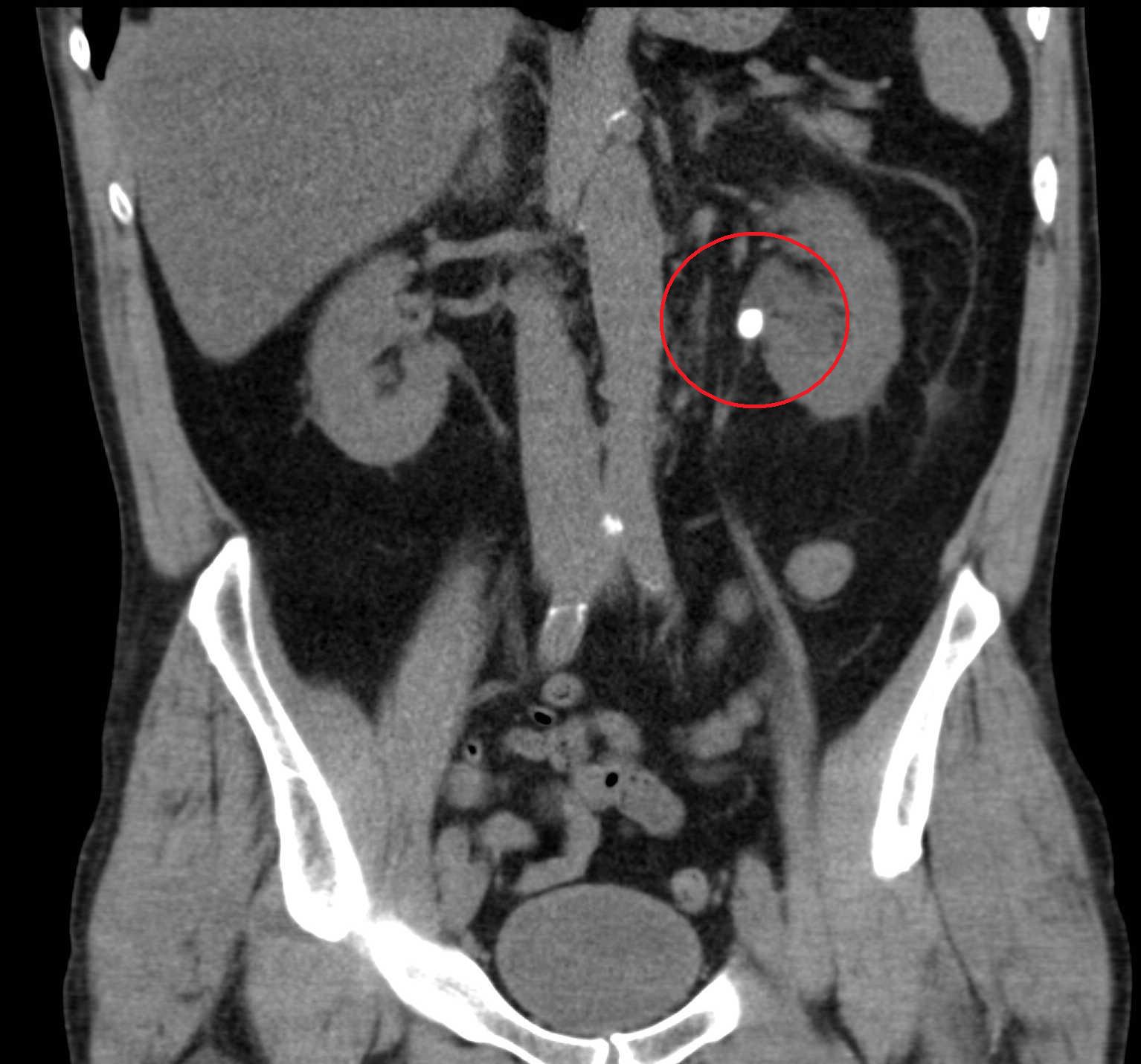 |
Urinalysis is usually done. It may show blood or pus in the urine whether or not symptoms are present.
For people with diagnosed stones, doctors often do tests to determine the type of stones. People should attempt to retrieve stones that are passed. They can retrieve stones by straining all urine. Stones found can be analyzed. Depending on the type of stone, urine tests and tests of blood chemistries and hormone levels may be necessary.


