Towards a Bi-Directional Neural Prostheses for the Central Nervous System |
||||||
 |
Current Status of Bi-Directional Neural Prostheses |
|||||
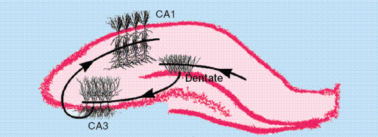
Hippocampus: The Current Central Nervous System Region of InterestCurrently the hippocampal region of the brain is the subject of the majority of research for bi-directional central nervous system prostheses. Furthermore, the hippocampus is a frequency site of damage during neurological diseases such as epilepsy, stroke as well as Alzheimer's disease [2]. Therefore, the creation of a neural prosthesis that mimics the functions of the hippocampus would have a large impact on the quality of life of many patients. Step 1:The hippocampus is made up of 3 sub-regions. These sub-regions are the dentate, CA3 and the CA1 that are organized into a excitatory cascade circuit of the form dentate -> CA3 -> CA1 [2]. It is possible to preserve the cascade network of these 3 regions when creating hippocampal slices. Therefore, in order to create biomimetic prosthesis for the hippocampus this cascade network was analyzed for nonlinear dynamics with the aim of creating the prostheses system for the CA3 region [2]. The cascade is shown in the figure below [2].
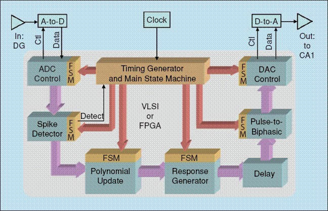
The nonlinearities of the CA3 region were analyzed by stimulating the afferents to the dentate with random impulse trains. These inputs to the afferents of the dentate resulted in nonlinear outputs that would eventually be the inputs to the CA3 regions. The nonlinear dynamics of these CA3 inputs to outputs was analyzed in this manner [2]. Step 2:The experimental data that was obtained from the experiments performed in Step 1 were used to estimate the Volterra-Poisson model for the CA3 region. Step 3:The model generated from Step 2 was used to implement a Field Programmable Gate Array (FPGA) based hippocampus CA3 region system that was capable of operating in real-time. A block diagram of the FPGA system developed is shown below [2].
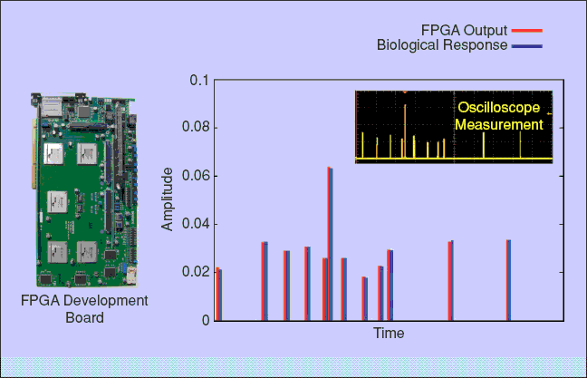
The response of the FPGA based hippocampus CA3 system was shown to correlate well with the observed biological response and this data is shown in the diagram below along with an image of the FPGA system [1].
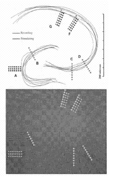
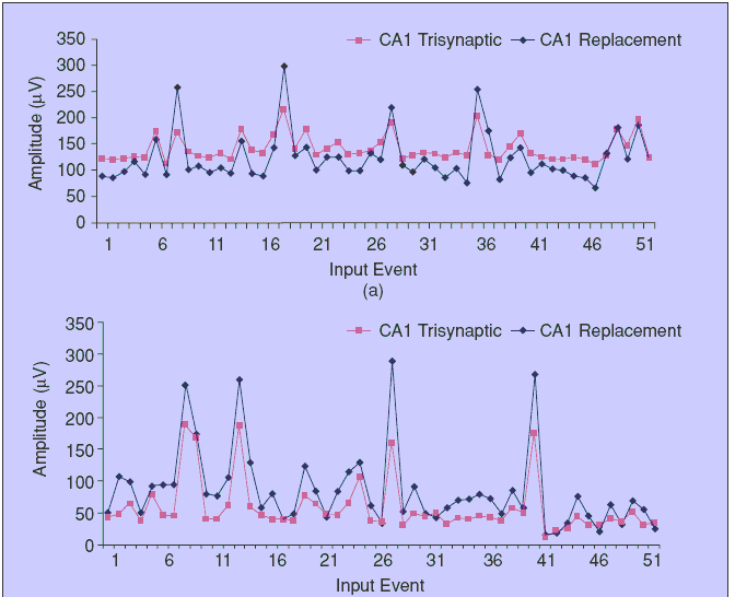
Step 4:The last step in the development of a FPGA based CA3 prostheses were to be able to interface the FPGA based system with the hippocampal slice and record the response of the system. The system was tested by means of comparing the CA1 output of a hippocampal slice when the biological CA3 region was replaced with the FPGA based model with the CA1 output of a hippocampal slice when the biological CA3 region was intact [2]. Below are images of the conformal electrode array utilized in these studies and the results that were obtained from the study [2].
| ||||||