![]()
![]()
![]()
![]()
![]()
3D STORM In order to resolve many of the cell's finer structures and organelles with far-field fluorescence, it is necessary to perform micoscopy in all three dimensions. Huang et al. have recently taken supperesolution imaging a step further to achieve three-dimensional stoichastic optical reconstruction microscopy (STORM) [6]. Superresolution techniques, such as 2D STORM have achieved impressive lateral resolutions upwards of 20-50 nm, and axial resolutions have been impoved by around 100 nm when implementing 4Pi and I^5M microscopy. The method takes advantage of the fact that the shape of image in the axial direction contains information about an objects position in that direction. By introducing a weak cylindrical lense into the optical pathway,the resulting astigmatism, or defocusing, causes a slight separation in x and y's focal planes. The fluorophore image now varies in the axial direction, where round indicates perfectly in between x and y (average focal plane with equal PSF widths), and elliptical shapes indicate some degree above (long axis on x) or below (long axis on y) the average focal plane. Images are fit with a 2D elliptical gaussian function to obtain x and y coordinants as well as peaks widths giving high accuracy positional information in all three dimensions. Figure 1. Left: Optical setup with cylindrical lens introducing the astigmtism. Right: x and y PSF widths at at varying focal planes. In a similar fashion to 2D STORM, reversible photoswitchable cyanine dyes were iteratively and stoichastically activated and deactivated to produce images with lateral resolution of 20-30 nm. In this case, activator-reporter pair, Cy3 and Alexa 647 respectively, were cyclically activated hundreds of times without photobleaching to obtain images which were subsequently averaged and fit to obtain positions. The efficiency of reactivation depends on the proximity of the activator dye. Calibration curves for cellular in vitro experiments was done by immobilizing 200 nm biotinylated polystyrene beads on a glass substrate, and subsequently incubating and thus coating the beads with Cy3/Alexa 647 labeled streptavidin. Figure 2. Conventional direct immunofluorescence imaging and 3D STORM of Left: microtubule network, and Right: CCPs in epithelial cells. Proof of concept experiements for 3D STORM were performed in BS-C-1 monkey kidney epithelial cells (see above). In the first study, the microtubules were immunolfluorescently stained and imaged. In a separate experiment also using immunfluorescence, Huang et al extended their application to resolving the three dimensional structure of cytosolic clathrin-coated pits (CCPs), which are spherical cage-like structures that participate in endocytosis.
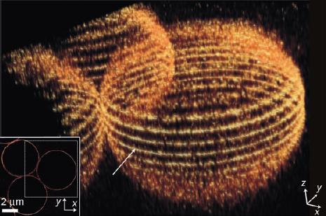
3D PALMIRA (PA-PALMIRA) Using a highly stable short-lived photoswitchable rhodamime derivative (SpaRh), Folling et al. were able to achieve PALM imaging in 3D with approximately 10 nm resolution [4]. The new reporter molecule has a high signal to noise ratio, simplifying image processing, obviating the need for TIRF illumination, and allowing use of higher densities per volume. Further implementation of two photon absorbtion (2PA) allows activation fluorescent states within a single focal plane of a thick sample. As a result, optical sectioning is achievable using picosecond pulses of red light followed by reconstruction via stacking fluorescently reconstructed images taken at different focal planes along the optical axis.
Figure 3. Reconstructed 3D PA-PALMIRA image of 5 um silica beads coated with SpaRh
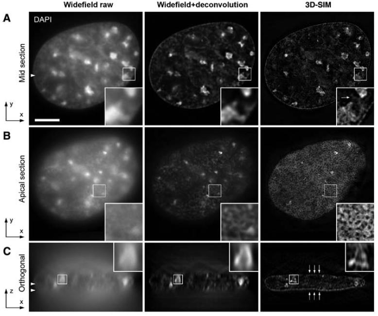
3D Structured Illumination Microscopy (3D-SIM) In a similar manner to STED, structured illumination requires making interference patterns on sample surfaces. Low-frequency moiré patterns result from an aliasing from harmonics in the linear combination of opposing high frequency tranverse waves. Mathematical techniques are used to form a real image at enhanced resolution from multiple individual moiré images acquired using different illumination pattern orientations. The resolution enhancement of structured illumination microscopy depends on the spatial frequencies contained in the illuminating pattern; however, the maximum frequency is still limited by diffraction and typically results in a resolution enhancement of no more than a factor of 2 (linear structured illumination). Sub-diffraction microscopy is achievable through either using photoswitchable probes as discussed earlier in other techniques, or by saturating fluorescent molecules to produce a nonlinear response where higher frequency hamonics increase imaging resolution. While in first generation SIM systems two beams were used to make an interference pattern, 3D-SIM utilizes 3 beams where illumination patterns can be controlled along the optical axis as well as laterally [7].
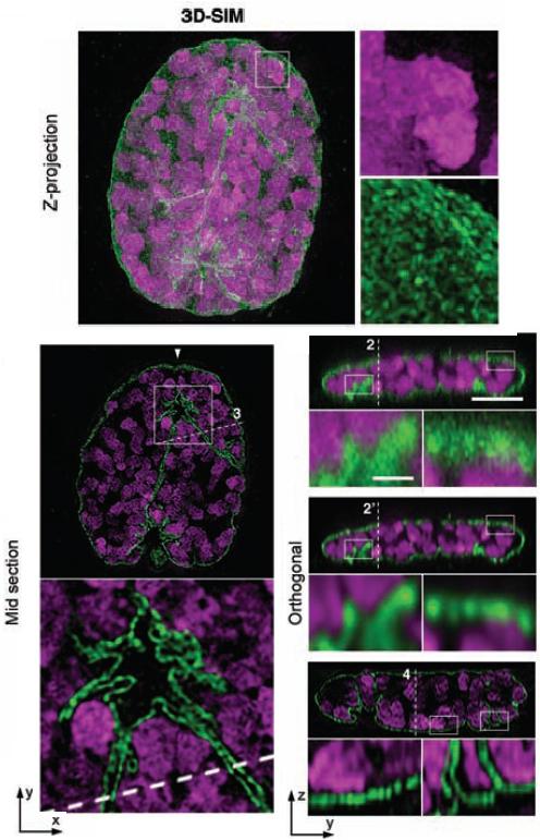
Figure 4. Left: Comparison of interface chromatin fine structure in DAPI stained C2C12 cells. Right: Different cross-section of immunostained invaginations in the mitotic envelope of C2C12 cells. Lamin-B is green and DAPI is magenta.
|