There have been many new technologies developed to acquire and process superresolution cellular and subcellular images. Below is an highlighted overview of the main technologies and how they work.
Stimulated Emission Depletion (STED) STED was one of the first superresolution methods presented to overcome the diffraction barrier of light. In this clever technique, a standing wave of intensity I(r) and wavelength λ is created at the specimen interface from the interference of two opposing light sources [Hell, toward]. Photoswitchable molecules within the specimen are either in a fluorescent absorbing state "A" or in a non-fluorescent non-absorbing state "B," and transition between the two states follows simple fist order kinetics with rate constants Kab and Kba. With a high enough light intensity, the interference pattern created on the surface of the specimen causes saturation, or stimulated depletion, of fluorophores everywhere (non-fluorescent state "B") except nodes where fluorescent molecules are unexposed to light. Therefore, the fluorescent signal is confined to nodes of the standing wave, and full fluorescent signal of the interrogated specimen can be mapped out by scanning the pattern on the sample. The minima of the standing wave f(x)=sin2(2πnx/λ) creates A with a full width-half max (FWHM) of dx=(λ/πn)arcsin(√(kBA/σImax)=λ/πn√ς, where the saturaion factor ς = Imax/Isat. Therefore the resolution of the nodes and thus the resolution for the detection signal is solely dependent on the saturation factor, ς, or the factor by which Imax surpasses the required intensity threashold for the photoswitchable fluorophores [2,3].
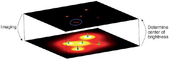
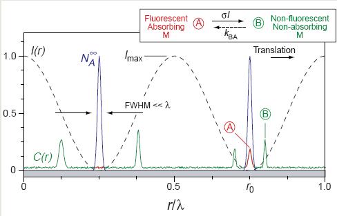
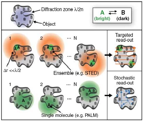
Figure 1. Fluorescent A molecules are detected at nodes of standing wave, while stimulated depletion occurs via saturation everywhere else with a high enough light intensity. Highest resolution of fluorescent signal is achieved via the combinatory effect of normal fluorescence, the STED node, and saturation depletion at that node.
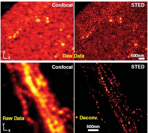
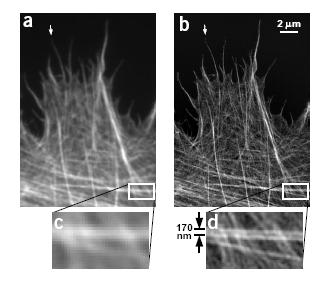
Figure 2. Left: Structured order of fused silica beads and neurofilaments are revealed in STED, but virtually indistinguishable in diffraction limited confocal microscopy. Right: SSIM reveals the fine architecture of microtubules that where standard epifluorescence microscopy loses the fine detail. STED and other similar methods such as ground state deplettion microscopy (GSD), and saturated pattern excitation microscopy (SPEM)/ saturated structured illumination microscopy (SSIM), that take advantage of reversible saturable or photoswitchable probles have been all classified under as reversible saturable optically linear fluorescence transitions (RESOLFT).
Photoactivated Localization Microscopy (PALM) / Stoichastic Optical Reconstruction Microscopy (STORM) PALM and STORM are two virtually identical techniques that instead of activating probes sequentially by scanning like RESOLFT microscopies, stoichastically activate probes within a sample (Figure 3, [2,3]). Sequentially acquired images of randomly activates reporter molecules are reconstructed into a single image thus separating fluorescent events that would have taken place within the diffraction limit. These techniques further recognize that since only localization and not the conformation of the molecular interaction is desired, unnecessarily large 3D fluorescent image structures can be represented by a single point to indicate coordinates of the fluorescent event. Centroids (center of mass of pixels) of fluorescent images are therefore simply fit with a scaled normal distribution, replacing complex pixel masses with a single point.
Figure 3. Activation scheme for activation of fluorescent molecules stoichastically (bottom: PALM/STORM) or by scanning (top: STED)
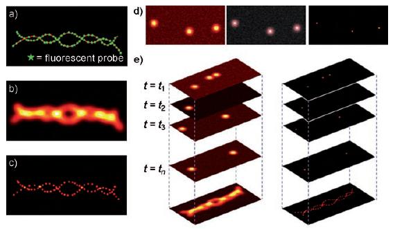
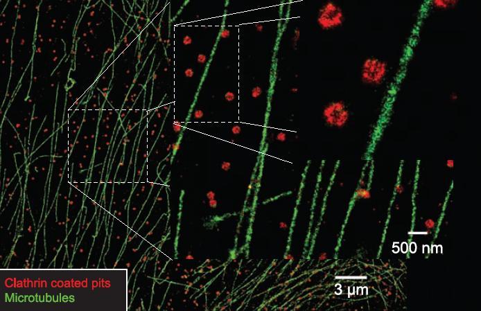
Figure 4. Left: Helical fluorescent probes are stoichastically activated/deactivated to acquire sum PALM image of the structure [5]. Right: STORM images of microtubules (green) and cytosolic clathrin coated pits (CCPs, red) that are thought to participate in endocytosis. Both: Nanometer localization is achieved by subsequent 2D gaussian fitting of fluoroscence images [6]. Unfortuntely, no microscopy technique is perfect, and PALM/STORM are certainly no exception. Due to the stoichastic nature of the technique, many activation cycles are needed for imaging large quantities of molecules, and this can lead to very long acquisitions times. Moreover, the 2D methods described here are only capable of nanometer resolution in the sample plane. The lack of optical sectioning capability means that samples have to either be very thin or illuminated via total internal reflection (TIRF). In order to overcome some of these limitations variations of the PALM technique have been created such as PALMIRA (PALM with independently running acquisition), which simultaneously implements two photon activation. See the "New Developments" section to take a look extension of PALM and STORM in 3D!
|