|
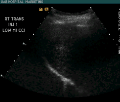
During the past 35 years, ultrasound and nuclear medicine were introduced into clinical medicine, computed tomography (CT) revolutionized diagnostic procedures and magnetic resonance imaging (MRI) emerged, bringing new diagnostic information at the cellular level. X-ray morphed from analog films to digital, and virtually all medical images became "soft" files on the electronic superhighway. Microbubbles have a high degree of echogenicity, which is the ability of an object to reflect the ultrasound waves. The echogenicity difference between the gas in the microbubbles and the soft tissue surroundings of the body is immense. Thus, ultrasonic imaging using microbubble contrast agents enhances the ultrasound backscatter, or reflection of the ultrasound waves, to produce a unique sonogram with increased contrast due to the high echogenicity difference. In this report, I’ll present a lipid shell-based PFC gas microbubbles demonstrates the potential of microfluidics as an efficient means for custom-designing contrast agents with different gas composition and new shell materials for stabilization. A nice prospect is the building of functionalized ‘smart’ microbubbles for targeted imaging and therapeutic applications such as localized drug delivery. |
|
Using Microbubbles for Improving Sensitivity in Molecular Imaging |
|
BME 240 Chia-Sheng Liao Spring 2008 |