|
Nearly 35 year ago, Gramaik and Shah (1968) discovered that the injection of microbubbles could produce the increase in ultrasound backscatter. They injected a dye into the heart through a small bore needle to create bubbles which were detectable on a M-mode echocardiogram. The use of these microbubble contrast agents has been shown to have significant clinical importance. Many contrast agents are either air-filled or contain gases that dissolve poorly in the blood with a mean diameter on the order of 1 to 10 μm such that they can reach the left ventricle of the heart and avoid rapid disappearance. The high echogenicity feature of microbubbles is useful in increasing the backscattered signal intensity (~15-20 dB) from blood. Increasing the echogenicity of blood allows clinicians to better measure blood perfusion in tissue and flow velocity in blood vessels, especially in recognizing malignant tumors or in monitoring treatment. |
|
Using Microbubbles for Improving Sensitivity in Molecular Imaging |
|
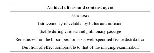
Contrast Agents |