E-mail:
Knowing exactly the ideal cell types that induce axonal regeneration is unknown, however, a number of studies have shown some success with certain cells. Currently, some suggested cell types include:
- Schwann Cells
- Stem Cells
Both cells have been shown to promote axonal regeneration in nerve guidance channels.
Recent studies have shown that Schwann cells, as seen in Figure 1, provide a favorable substrate for regenerating axons. Schwann cells release certain extracellular matrix (ECM) proteins, specifically laminin and collagen (click here for an overview of the ECM). Also, immediately after injury, Schwann cells form columns in which they express adhesion molecules for axonal lengthening. Furthermore, Schwann cells release growth factors that amplify nerve migration. These growth factors (click here for an overview of growth factors) include NGF, BDNF, and CNTF.3
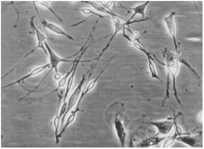
Figure 1. Schwann cells.3
As described earlier, Schwann cells are an important component of nerve guidance channels; however, possess difficult characteristics. For instance, purifying Schwann cells is very difficult. In addition, to ensure there is no immune response, the Schwann cells should be from the patient. These necessary procedures mean that culturing cells to prepare for transplantation can take as long as 10 weeks. Immediate surgery is required to restore most if not all function after nerve injury; as a result, this delay in time can drastically affect the benefits of nerve guidance channels.7
As a result, studies are gearing toward the use of stem cells as an indirect substitute for Schwann cells. Differentiating stem cells into Schwann cells eliminates the costs and immunology affects. Current stem cells capable of Schwann cell differentiation include neural progenitor cells, olfactory ensheathing cells, and mesenchymal-stem-cell-derived cells. As seen below in Figure 2, mesenchymal-stem-cell-derived cells are shown having a similar anatomy to Schwann cells (Figure 1).7
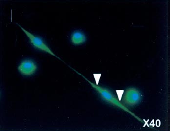
Figure 2. Mesenchymal stem cells.7