Peripheral Nerve Anatomy
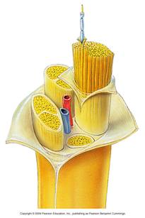
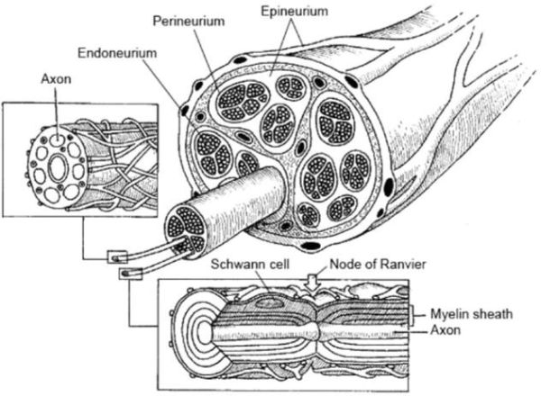
A peripheral nerve trunk such as the radial or medial nerve of the arm are comprised of axons of multiple neurons bundled in connective tissue fascicles surrounded by perineurium. Within the nerve, microvasculature runs along the outer layer (epineurium) with a transverse capillary network perfusing the endoneureum. Each fascile itself is comprised of endoneurium containing multiple neurons surrounded with myelin produced by Schwann cells. The soma and synaptic junction of the neuron cells is typically located at the basal ganglion roots of the spine with just the single axon traversing through the major length of the nerve trunk.
 
Peripheral nerve trunks are comprised of neurons traversing in fascicles within the body of a nerve. The perineurium is vascularized via microvasculature. ("Nerve Anatomy." A.D.A.M Anatomy. 2009)
Injury:
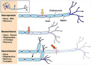
Several classification methods exist to describe the extent of injury, thereby determining regeneration potential, of peripheral nerve injury. Typically Seddon[4] or more complex Sunderland[5] classification schemes are used. Seddon classifies injuries into one of three categories:
Neurapraxia:
A low severity injury that typically leads to complete recovery. The structure of the nerve remains intact but electrical conduction down the axon is interrupted, typically by ischemia or compression injury, additionally secondary injuries can be caused by vascular damage leading to intrafascicular edema. The effects of this class of injury typically last from hours to weeks.
Axonotmesis:
Disruption of the neuronal axon takes place but the myelin sheath is still intact. Typically this is caused by a crush based injury, and not a laceration. If the neuronal tubules are maintained in place, regeneration and restoration of sensory or motor ability may return. Depending on the severity of the injury, regeneration may occur over the timescale of weeks to years.
Neurotmesis:
Characterized by not only loss of nerve conduction, but damage to surrounding nerve trunk connective tissue. In extreme cases of this injury category, complete transsection occurs, and commonly a neuroma forms over the proximal stump of the nerve, preventing normal continued regeneration to occur.

A graphical description of Seddon's classification of nerve injury as described for a single neuron.
Nerve Regeneration Process:
Upon injury due to small scale trauma, restoration of function begins to occur when the cause of the trauma is removed. Typically this is due to ischemia as a product of compression. Throughout this process, if the nerve tissue remains intact, the typical first step in the PNI regenerative process, Wallerian degeneration, does not occur. A more severe injury is one that damages the neuronal axon, yet maintains the myelin sheath. Once axonal continuity is lost, the process of Wallerian degradation occurs within 24 hours, causing the breakdown of the distal nerve stump. A the same process but to a lesser extent occurs at the end of the proximal nerve segment. Prior to the degradation process, the distal nerve component remains electrically active. Initially the axonal cell membrane degrades followed by the degradation of the neuron organelles and internal structures of the distal neuron. Secondarily, the myelin sheath is degraded as an action of the myelin producing Schwann cells. Macrophages are recruited to clear out cellular debris from the remaining neural tubule. Sprouting fibers from the proximal end of the severed neuron will grow down the neural tubules eventually reaching the motor end plates or sensory organ. This process is ultimately hindered if a severe misalignment or gap remains present or a neuroma scar blocks the end of the distal end. Surgical procedures are used to correct these issues so that proper regrowth occurs.
|