|
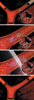
Figure 1 Microbubbles are a proprietary formulation of a lipid shell encapsulating an inert biocompatible gas. microbubbles allows them to penetrate a blood clot, so that when ultrasound is applied their expansion and contraction, or cavitation, can break the clot into very small particles. It is believed that these product candidates have the potential to treat a broad variety of vascular disorders associated with blood clots. |
|
Using Microbubbles for Improving Sensitivity in Molecular Imaging |
|
Result |
|
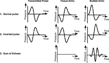
Figure 3 Graphs illustrate the principle of pulse inversion. A pulse of sound is transmitted into the body, and echoes are received from the contrast agent and tissue. A second pulse, an inverted version of the first, is then transmitted in the same direction, and the two echoes are summed. Linear echoes from tissue cancel each other. Nonlinear components of the microbubble echoes are reinforced when summed, producing a strong harmonic signal. |
|
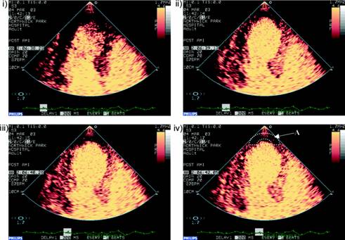
Figure 4 Contrast echocardiographic sequence with microbubble contrast agent defining myocardial perfusion within different myocardial segments i) is immediately following a high power ultrasound flash which destroys the micro-bubbles within the myocardium. ii)–iv) show replenishment of micro-bubbles in the septum and lateral walls within 2 heartbeats. A clear apical perfusion defect (A) is demonstrated which persists. |
|
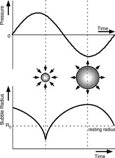
Figure 2 Graph illustrates microbubble behavior in an acoustic field. Bubbles respond asymmetrically to high-intensity sound waves, stiffening when compressed and yielding when expanded, a nonlinear response that produces harmonics in the scattered wave. |