3D Ultrasound

05/31/07

Home
Background
Screening Ultrasound
Probes
3D Ultrasound
Doppler Ultrasound
Future of Ultrasound


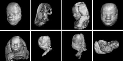
In the past two years, ultrasound machines capable of three-dimensional imaging have been developed. In these machines, several two-dimensional images are acquired by moving the probes across the body surface or rotating inserted probes. The two-dimensional scans are then combined by specialized computer software to form 3D images.


3D imaging allows you to get a better look at the organ being examined and is best used for:

bulletEarly detection of cancerous and benign tumors
bulletexamining the prostate gland for early detection of tumors
bulletlooking for masses in the colon and rectum
bulletdetecting breast lesions for possible biopsies
bulletVisualizing a fetus to assess its development, especially for observing abnormal development of the face and limbs
bulletVisualizing blood flow in various organs or a fetus


Home | Background | Screening Ultrasound | Probes | 3D Ultrasound | Doppler Ultrasound | Future of Ultrasound

This site was last updated 05/31/07