
![]()
![]()
![]()
![]()

![]()
![]()
![]()

Classic IVUS image
The typical image taken by IVUS system is a detailed image of the cross-section of the blood vessel, which it can distinguish the layers of catheter, lumen, intima, media, and adventitia. The first of the following figure is an IVUS images with normal or minimal disease. We can clearly see the geometry and morphology of different layers of blood vessel. This is because different layers have different composition of smooth muscle, collagen, and elastin, and thus its acoustic impedance is different.
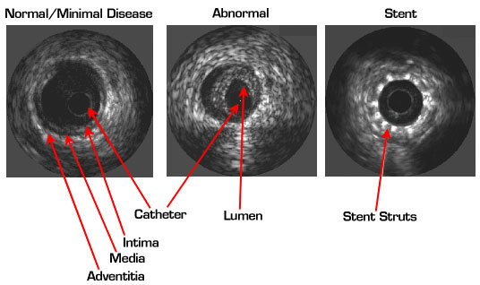
For an atherosclerotic vessel, plaque often forms in the layer of intima and occupy the lumen of the blood vessel. The second picture shows an example of this kind of abnormality, where the lumen of blood vessel is much smaller than normal. Because of the acoustic impedance of plaque is different with respect to its composition, IVUS possesses the unique capability to diagnose different kind of plaque (Link to Quantitative Measurement). Thus, IVUS becomes a gold standard technique for the detection and evaluation for coronary artery disease (CAD) and utilized adjunct to ballon angioplasty. The third IVUS image of the above pictures clearly shows the successfulness of implant a stent into the lumen with plaque. Again, this is because the stent itself has its unique acoustic impedance, and IVUS can decode its position via its acoustic reflection waves.