

Overview
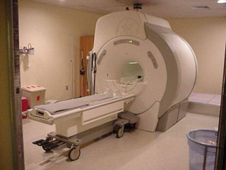
MRI has revolutionized clinical imaging, but several important challenges have limited it's application in the operating room. The first is the magnet itself. To do imaging, a large magnetic field is required. This, in turn, requires a large magnet that can be superconducting, permanent, or an electromagnet. In any case, the magnet takes a large amount of space and can interact in negative ways (like when the magnet sucks in an oxygen canister and crushes the patient). RF coils, gradient coils, and shim coils are also required hardware for MRI. These are more flexible than the magnet, but still require considerable space. Also, special tools have to be used that are MR compatible. |
 |
The main disadvantages of MRI are its cost, size, and potential harm to the patient. The advantages are that MRI uses low-energy, non-ionizing radiation that is relatively safe under prolonged exposure. This makes MRI a candidate for real-time, or near-real-time imaging. Also advantageous is the ability of MRI to distinguish different soft tissues. MRI has an obvious application for tumor removal surgeries. In a study where surgeons believed that had removed at least 90% of a tumor, post-surgical MRI revealed that only 50% of the tumor had been removed on average [Blanco, R.]. The result of incomplete tumor removal is, at best, return of the tumor, and, at worst, death. Any piece of tumor left in the body can quickly regrow so it is vital that tumors are completely (or as completely as possible) removed during surgery. |
|
Sources:
www.brighamandwomens.org
Blanco, R.; "Interventional and Intraoperative MRI at low field scanner--a review," European Journal of Radiology; 2005
Website created by: David Thayer, last edited: May 29, 2006