Piezoelectric
Materials Applications for Cardiovascular Devices
Website designed by: Luis
Alonzo
Artificial Heart Valve Sensor
Physicians often rely on sounds to be able to
assess the functionality of heart valves. The normal heart sound can be
typically described as “lub-dub”, and corresponds to the heart valves closing.
The “lub” sound is usually softer and last longer than the “dub” sound, and it
is associated with the closing of the atrio-ventricular (AV) valves at the
beginning of systole [9]. The “dub” sound corresponds to the closing of the
semilunar valves (aortic and pulmonary valves) [9]. The sound is shorter and
loader during this period due to the greater rigidity of the semilunar valves.
The following video shows what these sounds are like [10]:
By listening to the heart sounds the physician is
capable of assessing the conditions of the heart valves. Variations from the
normal “lub-dub” can indicate the presence of heart valve stenosis or insufficiency
[9].
Piezoelectric based sensors are being designed by
Hall et al., for the purpose of converting heart sounds into a visual and quantitative
tool [11]. This particular piezoelectric crystal displays a sensitivity of 10 mV/dyne
force and 40 mV/micron displacement, capacitance of 20 nF, and frequency
response of 0.02-2 kHz [11]. However,
one major problem is that the heart sound recording via this method is
overshadowed by unwanted background noise. The authors attribute this issue to
various sources, such as: contact between skin and stethoscope, breathing, and
talking during the procedure [11].
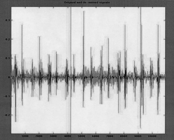
Moreover, an approach to
reduce this noise was investigated by this group. They utilized the method of “wavelet
denoising”, which relies on the wavelet transform rather than the Fourier
transform [11]. This method is used due to the similarity between the wavelets
and the time-domain shape of the heart sounds. Fortunately, as shown in the
figure below, this technique shows that the noise can be greatly reduced by
applying a particular threshold [11].
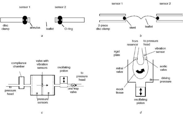
A new approach by Lanning
S. and Shandas R. uses piezoelectric sensors mounted on artificial heart valves
to analyze their functionality [12]. The sensor is a noninvasive method used to
detect variations normal heart valve vibrations. The loading of thrombi or irregular
vibrations will create a heart signal which can indicate forthcoming
malfunction. The figure below shows several diagrams of the piezoelectric
sensors mounted on tww different heart valve models [12]:
Results of this research [12] showed that the frequency content of the
sounds generated by the mechanical valve closing (first model) contain peaks
between 100Hz-10kHz, while the closing sound of the bioprosthetic valve (second
model) contain a lower frequency range. In addition, the sensors were able to
detect leaflet stiffening; showing and increase in the frequency peak from 474+15Hz
to 1154+12Hz. Finally, thrombus formation was also successfully measured
through this method; showing a decreased in the total low frequency range as the
thrombus formation increased.