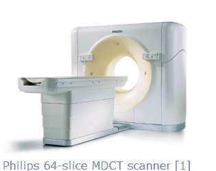
The first 64-slice multidetector computer tomography scanner reached the US in 2005. The MDCT is a revolutionary imaging technique which decreases scanning time without compromising image quality. A scan of a 30cm section of the body can be completed in 6 seconds, which allows for increased x-ray power and smaller radiation dosage. The MDCT can accurately and automaticaly determine the x-ray dose for each slice of a patient’s body. For instance, higher dosage is used for the shoulders and the pelvis, and lower dosage is used in the abdomen. [2] The software package can easily compile the image slices into a detailed 3D view, potentially changing the way radiologists view CT scans.
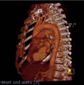 Common uses of the MDCT include diagnosing cancers, scanning the chest and abdomen, evaluating spinal injuries, and detecting and treating vascular diseases. Arguably, the 64-slice MDCT can rival the angiogram in visualizing the coronary arteries because it is faster, safer, noninvasive, and less expensive. [1] The MDCT can be used for detailed soft tissue studies with the correct application of contrast media. Intravenous contrast media improves the opacity of soft tissues, lesions, and blood vessels. High resolution reconstructions of brain matter, sinuses, and inner ear studies can easily be performed.
The speed, image resolution, z-axis coverage, and real-time capabilities of the 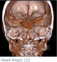 MDCT may enable new diagnostic and therapeutic techniques. The capabilities of the MDCT can be used throughout the planning, targeting, and monitoring stages of a complicated procedure. [3] Research is ongoing to combine the MDCT with many interventional oncology treatments.
One of the newest interventional oncology techniques is percutaneous radio frequency ablation. RFA is predominantly used to treat smaller tumors, less than 5cm, when surgical removal is not an option. During the procedure, needle-like RFA electrodes are inserted into the tumor. The high frequency alternating current then increases the temperature inside the tumor, killing the cells. Currently, the preferred method to guide and place the electrodes is ultrasound because of its real-time capability, visualization of nearby vasculature, and high speed. [4] With the benefits of MDCT, researchers are hoping to increase the accuracy of the procedure, minimize the risk of damage to surrounding tissue, and enable the treatment of larger tumors.