Multilayer Nanoencapsulation
---------- New Approach for Immune Protection of Human Pancreatic Islets
![]()
![]()
![]()
![]()
![]()
![]()
![]()
First an encapsulation protocol for the Langerhans islets was developed. After islet encapsulation, their functionality was characterized by release of insulin, sensitivity to stimulation intensity by static glucose stimulation, and long-term viability and functionality of the islets. Furthermore it was possible to show that the capsule prevents the recognition of the islets by antibodies. Several polyelectrolytes couples were tested for their suitability. From the results it can be concluded that the nanoencapsulation of pancreatic islets is a promising alternative to the commonly used microencapsulati on with ultrapure alginate beads. In the beginning it was necessary to investigate how cells were stressed by the encapsulation process itself with its repeated medium changes and if the cells survive the encapsulation process. Then it was also interesting to determine the maximum number of layers which can be applied without causing too much damage to the islets. Different polycations were used as first layer because from previous investigations it was known that cells have different sensitivities to diverse polyelectrolytes.
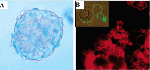
(A) Light
microscopy image of trypan blue-stained islets after encapsulation with three
pairs polyelectrolyte layers (PDADMAC/ PSS)3. The dead cells appear dark blue.
(B) Fluorescence microscopy of the coated islets. Only imaging of the border
region was possible due to the thickness of the islet. The applied PAH layer
appears red due to covalently bound Alexa-555. The polymer penetrates also the
intercellular space (bright red). Inset: Smaller fragment of an pancreatic
islet. The merged images of transmission and fluorescence microscopy that the
capsule surrounds the cells. Here the fluorescence is due to green-fluorescent
FITC-PAH.
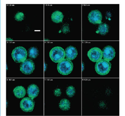
3D optical sections of encapsulated cells after incubation in DAPI. The homogeneous FITC-PATH green fluorescence is indicative of a uniform cell coating while the DAPI blue fluorescence, staining the mitochondrial and nuclear DNA. is indicative of the integrity of the main cell biostructures. Scale bar:2um.
![]()
Designed by Shan Yang