Optical Coherence Tomography (OCT)
For Imaging Vulnerable Plaques
![]()
![]()
![]()
![]()
![]()
![]()
![]()
 Basic Principles
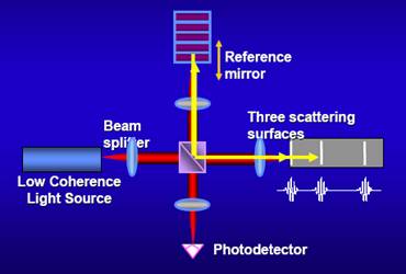
The principle OCT is white light or low coherence interferometry. The optical setup typically consists of an
interferometer with a low coherence, broad bandwidth light
source. Light is split into and recombined from reference and
sample arm, respectively.
In time domain OCT the pathlength of the reference arm is
translated longitudinally in time. The interference of two
partially coherent light beams can be expressed as
![]()
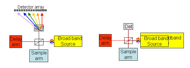
In frequency domain OCT the broadband intereference is acquired with spectrally separated detectors (either by
encoding the optical frequency in time with a spectrally scanning source or with a dispersive detector, like a
grating and a linear detector array).
Focusing the light beam to a point on the surface of the sample under test, and recombining the reflected light
with the reference will yield an interferogram with sample information corresponding to a single A-scan (Z axis
only). Scanning of the sample can be accomplished by either scanning the light on the sample, or by moving
the sample under test.
Designed by Jingjing Jiang