
Possibilities for Non-Invasive Methods of Detection.
The non-invasive detection of Hematocrit is an active area of research. There have been many technologies used to attempt to realize this goal. Ultrasound has been used but it was found that temperature variations introduced too much error. Doppler Ultrasound was used successfully on the brachial artery but excessive movement of the artery caused the readings to vary. NIR optical imaging also held promise but without direct access to the blood vessels, variations in the scattering and absorption properties of skin made NIR problematic. A review of the paper, "Toward noninvasive measurement of blood hematocrit using spectral domain low coherence interferometry and retinal tracking" by Nicusor Ifrimia et al (1), will be given as a current example of optical technology being researched that holds the promise of success. This paper can be found here.
The researchers show that the non-invasive determination of Hematocrit is possible using low coherence interferometry (LCI) combined with direct access to the vessels of the retina. The hematocrit information is derived from the slope of the LCI depth reflectivity profile.
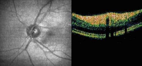
The researchers decide to use spectral-domain LCI (SDLCI) because of faster acquisition rates and far greater sensitivity over time-domain LCI. SDLCI has been used in ophthalmology to image the retina, measure Doppler flow in retinal vessels, and for spectroscopic measurements. However, eye movements make collection of data problematic. The beam interrogating the eye must be stabilized on a retinal vessel to collect depth-reflectivity profiles in the blood. The researchers use a combination of two technologies, direct retinal tracking with tracking scanning laser ophthalmoscopy (TSLO) and SDLCI, to measure Hematocrit non-invasively. Their preliminary measurements correlate well with conventional clinical methods.
The researchers demonstrate that reliable and quick non-invasive measurements of Hematocrit using SDLCI and TLSO are possible. However, calibration methods must be improved and larger studies done. They predict that refinement of the methods and instrumentation could lead to the development of a clinical tool and that this instrument could be used in a wide variety of physiological monitoring applications. (see paper for details on experimental setup and data analysis)