Anatomy of the Eye

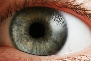
Images of the eye and its anatomy are shown in the slideshow at the bottom of this page. Please feel free to familiarized yourself with the eye’s basic structures as needed to gain a better perspective on the anatomical barriers to opthalmic drug deliver.
Many hard-to-treat eye disorders occur in the inner-ocular structures of the eye. Some of the most challenging areas to reach are the macula, aqueous humor, extravascular space of the retina and the vitreous body. Topical treatments or eye drops do not allow for sufficient diffusion in the anterior segments of the eye. [4] Systematically administered drugs must be administered in excessive concentration to overcome the blood-aqueous barrier or blood-retina barrier and typically cause adverse side effects. The blood-aqueous barrier is “a selectively permeable barrier between the capillary bed in the processes of the ciliary body and the aqueous humor in the anterior chamber of the eye” [3]. Similarly, the blood-retina barrier inhibits the perfusion of therapeutic drugs from the capillary bed into the retina [3].
To this extent a variety of alternative routes for drug deliver have been developed including intravitreal, intrascleral, and trans-scleral injections. Ultrasound targeted microbubble destruction (UTMD) would be delivered by these alternative routes, essentially sidestepping the barriers of current drug delivery systems. Microbubble colloidal particles would be loaded into the eye via subcutaneous injections. Then, microbubble cavitation from applied ultrasound (US) releases the drug payload and creates pores in the local tissue for drug uptake (see Microbubbles section) [4].
Barriers to Ophthalmic Drug Delivery