|
Introduction
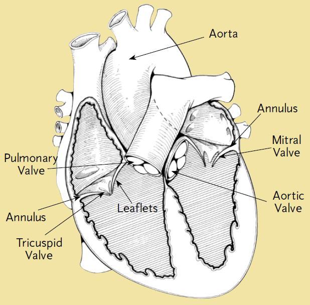
Blood is pumped through the chambers of the heart, aided by four heart valves. The valves are comprised of leaflets that open and close to let the blood flow in only one direction and the annulus that anchors the leaflets to the walls of the heart. When any of these extremely important structures become dysfunctional, serious and often life-threatening symptoms can occur.
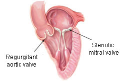
Valvular regurgitation and stenosis are the two main effects of valvular disease. Regurgitation, or insufficiency occurs when blood flows in the wrong direction as the leaflets close or after they are closed. This is a serious problem because it forces the heart to work much harder than normal to pump the same amount of blood. Stenosis is the condition when the valve opening narrows, reducing the amount of blood that flows through the valve while it is open. Stenotic valves also cause the heart to work harder, in addition to increasing the chances of blood clots due to the high shear forces on the blood as it is forced through the smaller-than-normal opening. See the pictures and video below for an explanation of these issues.

Symptoms
Patients with these effects often encounter the following symptoms:
- Shortness of breath, especially during or after activity or when lying down flat in bed.
- Recurring dizziness or weakness, even when performing normal activities.
- Pressure or heaviness in chest, especially during or after activity or when in cold air.
- Heart palpitations or irregular heartbeat, feelings of the heart skipping beats, or flip-flopping in chest.
- Swelling in ankles, feet, or belly.
- Sudden weight gain with possibly as much as 2 to 3 pounds in 1 day.
Diagnosis
Methods of diagnosis include the following:
- Electrocardiogram: An electrocardiogram is used to record the heart’s natural electrical currents, helping to analyze the heart's rhythm, rate, and the strength and timing of the electrical currents. This helps doctors uncover a patient’s underlying symptoms that may be signs of heart disease.
- Chest x-rays: X-rays are used to diagnose heart diseases and to evaluate placement of tubes, catheters, pacemakers, and defibrillators.
- Nuclear scanning: Nuclear scanning refers to imaging the body’s internal organs using radioactive waves, ultrasound or magnetic fields and helps in ways similar to x-rays.
- Echocardiography: Echocardiography is performed as either a transthoracic or transesophageal exam depending on the patient's particular circumstances. These tests use ultrasound to pick up sound waves that are moving through the heart. The waves are then converted into moving pictures.
- Cardiac catheterization: Cardiac catheterization is used to determine if a patient has coronary artery disease. It can also pinpoint any plaque build-up from atherosclerosis. This is considered to be the gold standard of imaging for determining the nature of coronary artery disease, but it is more invasive than other procedures.
|