Neurogenesis Background

Until recently, it was believed that neurogenesis only occurred in developing organisms. There has now been a paradigm shift and new research has shown that neurogenesis occurs throughout adulthood. In 1983, Goldman and Nottebohm showed neurogenesis in adult canaries. Eriksson et al. and Gould et al. showed that neurogenesis also occurs in mammals, including humans. Similar to blood vessel growth, new nerves can only grow from existing nerves.
After injury, neurons in the peripheral nervous system are capable of regenerating axons to reinnervate targets. There are a variety of criteria that must be met for peripheral nerves to repair injury. Neuronal survival after the injury is a prerequisite for regeneration. The axotomized neurons must convert from transmitting mode to a growth mode and express growth proteins, such as GAP-43, tubulin, and actin, as well as neuropeptides and cytokines. The sprouts must then reach the distal nerve stump of the severed nerve at the right time. At this point, Schwann cells undergo proliferation. This process takes time because the rate of axonal transport is slow. The Schwann cells must remain in the growth supporting mode for a long period of time. This process is inefficient and therefore recovery times are very long. New techniques are being developed to decrease the time it takes for recovery.
Axonal sprouting generally occurs when there is a loss of motorneurons due to trauma. It is a process in which nerves sprout from intact axons at either the motor endplates to give rise to ultraterminal sprouts, or the nerve terminals to give rise to preterminal sprouts, or to the nodes of Ranvier to give rise to nodal sprouts. Figure 1 shows the three previously discussed types of axonal sprouts.
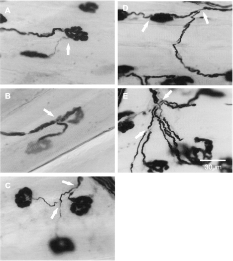
Figure 1. Three different types of axonal sprouts commonly seen in partially denervated muscles. Panel A, B and C respectively show ultraterminal, preterminal and nodal sprouts as indicated by the arrows. In extensively denervated muscles, more complicated sprouting was observed: Panel D) 2 types of axonal sprouts (axonal and ultraterminal sprouts, indicated by arrows) emerged from a single axon; Panel E) multiple axonal sprouts (pointed by arrows) emerged from the same axon.
Crisci and Ferreira found that by applying ultrasound with a frequency of 1.5 MHz and an intensity of 16 mW/cm (SATA), they could accelerate the regeneration of the sciatic nerve after neurotomy in rats. Mourad et al. used ultrasound with a frequency of 2.25 MHz and an intensity of 0.25 W/cm2 to produce the best recovery after sciatic nerve crushing in adult rats.