BME 240 Spring 2009
Matthew Kornswiet
Intraoperative Computed Tomography


Intraoperative computed tomography is an emerging field that strives to make CT scanning portable for both the operating room and intensive care situations. A variety of systems are being developed, three of which show the most promise in being able to reproduce a normal CT scan. While these three systems are relatively new and need to be tested more completely, they represent the future of intraoperative image guidance.
NeuroLogica CareTom Mobile CT Unit
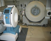
The NeuroLogica CareTom Mobile CT Unit, from the Massachusetts General Hospital and the Harvard Medical School, is designed so that the patient remains stationary and a gantry moves the machine to the next location. This setup makes it possible to CT a patient from a bed where he or she cannot be moved, such as in the operating room. The scanner itself has wheels and runs on power from an ordinary wall socket.
Figure 1: NeuroLogica CareTom Mobile CT Unit
In initial studies, the NeuroLogic CareTom showd a spatial resolution of 0.8 mm and a signal to noise standard deviation of 0.37 percent.
Figure 2:
Left set of 3: A pre-operative CT scan
Right: A intraoperative CT scan using the NeuroLogica CareTom Mobile CT Unit
Since the machine is mobile, it is possible for a hospital to share its machines (similar to the way current intraoperative x-ray machines are used) with many different operating rooms. Some restraints are that the patient has to be on a radiolucent bed, which is standard in x-ray cases, and that it takes time for the gantry to move. In addition, the patient is exposed to a higher dose of radiation than a current intraoperative x-ray and only the patient’s head can be scanned by the machine.
BrainSuite iCT
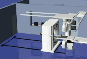
The BrainSuite iCT by Siemens and BrainLab is the size of a normal CT machine and therefore has the same resolution and the ability to scan the entire body. The machine is placed on tracks in the room and slides over the patient as needed.
Figure 3: The Brain Suite iCT
A limiting aspect of this design is that a patient in the operating room has many wires and other tubes coming from him or her, making it difficult to slide a machine over them. Since this machine is similar to a typical CT scanner, it produces similar amounts of radiation during scanning. In other words, the patient is exposed to a higher dose of radiation than that of a typical intraoperative x-ray, but a similar dose to that of a typical stationary CT scanner. While the BrainSuite iCT is still in production, it has potential to make Computed Tomography able to be intraoperatively done.
Xoran xCAT ENT Compact CT Scanner
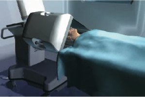
Finally, there is the Xoran xCAT ENT compact CT scanner by Xoran and Medtronic. This machine is the most portable of the three, but is limited to head scans.
Figure 4: The Xoran xCAT ENT compact CT scanner
Not only is it able to do a very detailed CT scan, 0.4 mm slices, but it also is able to immediately reproduce a three-dimensional image to be used by an image guidance machine. According to its specifications, it only takes 60 seconds to reproduce 300 transverse slices. In other words, it takes one minute to reproduce 24 mm of three-dimensional data when using the highest quality setting.
Figure 5: A intraoperative image and rendering taken using the Xoran xCat ENT compact CT scanner
The Xoran xCAT ENT runs on a normal wall outlet and has a very short scan time--20 seconds for a sinus scan. The short scan time minimizes the radiation exposure dose to the patient. In fact, there are very low doses of radiation as compared to the previous two systems.