BME 240 Spring 2009
Matthew Kornswiet
Computed Tomography Overview


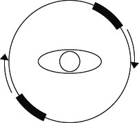
Computed Tomography (CT) scanning is a medical imaging technique that uses tomography to view a three-dimensional structure. The technique uses x-rays from multiple angles to visualize the structure. Conventional CT machines have an array of x-ray sources and an array of x-ray detectors arranged 180 degrees from each other with the patient in the middle.
Figure 1: A transverse cross section of a normal CT scanner
Both the detector and source array rotates around the patient 360 degrees. From there, the patient is moved so that the arrays translate down the vertical axis of the patient.
Figure 2: A longitudinal cross section of a normal CT scanner
This process creates multiple transverse cross sections that when put together create a three-dimensional image. Each x-ray beam travels through the body and is detected by the detector array. The body absorbs a certain amount of the x-ray beam and reduces the energy read by the detector. By assuming the ray travels in a strait line through the body, the detector reads the output as a line integral where the radiographic densities are added up along the path of the ray. The detector reads the intensity in Hounsfield units, which compares the density to that of distilled water.
For the first part of the imaging technique, rotating the source and detector arrays around the patient, gives a two dimensional image. The Radon transform is used to collect the information along the individual line integrals. The inverse Radon transform puts the image back together again by assuming that the intensity is split evenly over the width of the image. In other words, each pixel is made up of the average intensity from all of the detectors at that intersect that point.
Figure3:
Left: An example of a Radon transform being done on an image. The outer bars on four sides represent detectors.
Right: An example of an inverse Radon transform reconstructing an image. The outer bars on the four sides are the detectors.
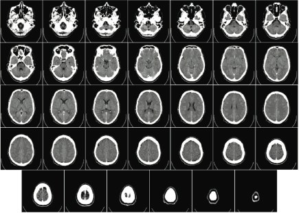
The final result is an array of two-dimensional cross sections that span the area of interest.
Figure 4: An array of two-dimensional cross sections produced by a computed tomography scan
Computed tomography is more comprehensive than a standard x-ray because it has the capability to produce a three-dimensional image and can take more images in a shorter period of time. However, a CT scan exposes the patient to a much larger dose of radiation than a normal x-ray and this increase in radiation needs to be considered.


