Background knowledge of Cochlea

Auditory pathway

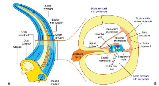
Cochlea is a snail-like structure in the inner ear. When sounds are collected from the pinna, through ear canal, then reach on the ear drum. The vibration of ear drum causes the three bones of the middle ear to vibrate and then the foot plate of the stapes vibrates in the oval window. Vibration of the foot plate causes the perilymph in the scala vestibuli to vibrate which in advanced induces the displacement of the basilar membrane. (Panel A in figure 1) Sounds with higher frequencies cause displacement of the basilar membrane near the oval window, and sounds with lower frequencies cause displacement of the basilar membrane some distance from the oval window. Movement of the basilar membrane is detected in the hair cells of the spiral organ, which are attached to the basilar membrane. (See panel A in Figure 1) Vibration of the perilymph in the scala vestibuli and of the endolymph in the cochlear duct are transferred to the perilymph of the scala tympani and then transferred to the round window, where the sound energy is dampened.

In this pathway, the outer hair cells (OHC) undergo nonlinear amplification of  the input vibration, and the inner hair cells (IHC) convert the amplified mechanical vibration via ion channels into electrical output which stimulates spiral ganglion neurons (SGNs) nearby. The electrical stimulus is then transmitted through the nerves to the brain. The cochlea has a tonotopic organization due to the numerous gradations along the length of the cochlea, such as basilar membrane (BM) stiffness (stiffer and short at the basal end), stereocilia length, hair cell somata size, SGN somata size, fiber length, and axon diameter [1]. The tonotopic frequency distribution from base to apex of the cochlea is from high to low.

Figure 1 The cochlea of the mammalian inner ear

(A) The cochlea. It is the stretched version of cochlea and modified in order to understand the sound transmission 

(B) Cross section of the cochlea. The cochlear duct is inserted in the perilymph. It is filled with endolymph and contains the organ of Corti between the tectorial and the basilar membranes.

Auditory cells

There are two types of cells in the organ of Corti: support cells and hair cells. The hair cells are "receptor" cells -- the ones that transduce sound. Support cells such as Deiter's cells support hair cells. The tops of the hair cells and pillar cells form the reticular lamina, which isolates the hair cells's stereocilia from their cell bodies. The tectorial membrane is loosely coupled to the reticular lamina. (see figure 2 below)

There are four rows of hair cells: one on the inner (modiolar) side of the tunnel formed by the pillar cells -- inner hair cells, and three on the outer side of the Tunnel of Corti -- outer hair cells. Deriter's cells support the outer hair cells at their base, and the walls of outer hair cells are surrounded by fluid. As for the inner hair cells, they are supported by the surrounded support cells.

Auditory nerve fibers also play an important role in auditory pathway. They provide synaptic connections between the hair cells of the cochlea and the cochlear nucleus within the brainstem. The cell bodies of the cochlear nerve lie within the central aspect of the cochlea and are collectively known as the spiral ganglion. This name reflects the fact that the cell bodies, considered as a unit, has a spiral shape.

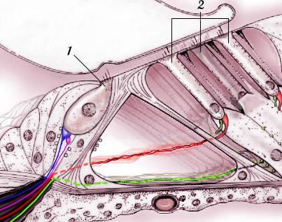
Type I neurons make up 90-95% of the neurons and innervate the inner hair cells. They have relatively large diameter, and are bipolar and myelinated. Each type I axon innervates only a single hair cell, but each hair cell directs its output to an average of 10 nerve fibers. Type II neurons, which ahve a relatively small diameter, connect with the outer hair cells, are monopolar and are not myelinated.

The transmission between the inner hair cells and the neurons is chemical, using glutamate as a neurotransmitter.
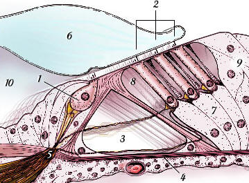
1-Inner hair cell,

2-Outer hair cells,

3-Tunnel of Corti,

4-Basilar membrane,

5-Reticular lamina,
6-Tectorial membrane,

7-Deiters' cells,

8-Space of Nuel,

9-Hensen's cells,

10-Inner spiral sulcus

Type I neurons: The radial afferents (blue) and the lateral efferents (pink) innervate the inner hair cells;

Type  II neurons: The spiral afferents (green) and the media; efferents (red) innervate the outer hair cells

Figure 2 Hair cells and supporting cells in the inner ear

Figure 3 Type I and type II neurons

Reference

[1] Davis, R. L., "Gradients of neurotrophins, ion channels and tuning in the cochlea", Neuroscientist, Volume 9, Number 5, 2003