University of California, Irvine: Biomedical Engineering (BME) 240 | Presented by: Katherine Lee

Designing Dendrimers for Biomedical Applications
Introduction
Drug Delivery
Therapeutics
Imaging
Tissue Engineering

Introduction

A dendrimer consists of a core, repeated iterations surrounding the core called dendrons, and the periphery groups which can be modified for ligand attachment.  The number of branches emanating from the core can be counted as subsequent “generations.”  The chemistries of the dendrimers vary greatly to affect solubility, degradability, and biological activity; however, some of the more common building blocks used are polyamidoamines (PAMAM), polyamines, polyamides, poly (aryl esters), polyesters, carbohydrates, and DNA.  Dendrimers have a wide variety of applications, including drug delivery vesicles, therapeutic agents, imaging contrast agents, scaffold materials for tissue engineering, and artificial enzymes.  The utility of dendrimers stems from the capability of tuning dendrimer characteristics, such as size, shape, and composition and the synthesis of dendrimers in a highly reproducible and consistent fashion.


A useful property of dendrimers is that as the generation number increases, the dendrimer size increases and the terminal groups become more tightly packed together to regulate release rates from the dendrimer interior.  The increased number of terminal groups allows for multiple ligand attachment sites and increases the probability of an affinity interaction.  Also, the terminal groups are important because they can be hydrophobic or hydrophilic, anionic or cationic, all which determine its interactions in the solvent.


Back to top

Dendrimer attachment to cellular membrane

Dendrimer conjugation to DNA strand


Drug Delivery

Dendrimers are used for drug delivery because the carrier can improve solubility, increase circulation half-life, and improve drug transit across biological barriers.  Polyamidoamine (PAMAM) dendrimers have carried the antitumor drug methotrexate and fluorescein for tracking [1].  The peripheral amines were coated with acetyl groups to reduce charge at the dendrimer surface.  The acetylated dendrimer was derivatized with folate as the target ligand to attach to the overexpressed folate receptors in KB tumors.  The concentration of the dendrimer was 5-10 times higher than the control group with dendrimers without folate ligands.  In mice, treatment with 15 biweekly intravenous injections showed reduced tumor growth rate when compared to mice treated with saline.  The diameter of the dendrimers was less than 5 nm, which means that the drug could be quickly eliminated by the renal system.  This results in a double-edged sword of reduced toxicity concomitant with reduced drug efficacy. 


Gene therapy could also benefit from the use of positively charged dendrimers complexed to negatively charged DNA.  Studies have shown reduced toxicity in transfected cells when compared to traditional polyamine agents [2].  However, to be useful in clinical applications, the issues surrounding the use of a positively charged carrier must be addressed because cationic substances are associated with toxic and hemolytic effects.


In drug-delivery, issues of biocompatibility often need to be considered and are characterized by retention levels in the body.  The ideal dendrimer must have a low molecular weight to be easily filtered by the kidneys.  The concept of enhanced permeation-and-retention effect is used to construct a mathematical model of tumor-targeting efficiency.  Important parameters for the mass balance of drug concentration are k-elimination (elimination of drug by kidneys), k-extravasation(dependent on tumor characteristics), k-release (dependent on chemistry of drug attachment to dendrimer and local environment), and k-washout(rate of drug elimination from the tumor) [3].  It is evident from these parameters that variations in dendrimer characteristics can affect drug release and tumor drug uptake.

Back to top

Therapeutics

While dendrimers can be used as drug carriers, they themselves can be used as therapeutic agents.  One group exploited the branching properties of the dendrimers to induce removal of prion proteins in infected cells, which proved to be more effective than linear or small polyamines [4].  The multivalency of surface ligands on dendrimers was further used to demonstrate that this property could inhibit foreign agents, such as bacteria, viruses, and proteins from binding to the cell.  Another study showed a G-4 poly(L-lysine) dendrimer with sulfate groups at the surface could bind electrostatically to viral envelope proteins  and block viral entry into the cell [5].

Back to top


Imaging

Dendrimers have been used as MRI contrast agent and oxygen-tumor sensing agents.  In tumors, the oxygen levels can be a measure or how well the tumor will respond to treatment.  Consequently, dendrimers made of poly(glutamic acid), poly(aryl esther) or poly(ether amide) can be encapsulated with metallopolyphorins.  These dendrimers are water-soluble, and the metallopolyphorins’ phosphorescence is quenched upon collision with oxygen [6,7].  In vivo or in vitro measurements can be made by phosphorescence excitation with visible or IR light. 


Dendrimers have also been recently used in the functional magnetic resonance imaging of the kidney which uses low-molecular weight contrast agents that can be filtered at the glomerulus but not absorbed or secreted by the tubules.  Gadolinium-bound dendrimers are used in specific renal parenchymal diseases and its uptake is indicative of damage to the proximal straight tubule in the outer medulla [8].

Back to top

Tissue Engineering

The peripheral functional groups in dendrimers can be modified to cross-link with one another to form an insoluble support structure.  Grinstaff et. al have employed dendrimers as injectable sealants in corneal wounds [9].  The study also showed the ability for the sealant to maintain its structural integrity and withstand maximum intraocular pressures.  Sontjens et. al. used a hydrogel that consisted of a polyethylene glycol core and methacrylated poly(glycerol succinic acid) dendrimer terminal blocks [10].   The scaffold was used to support chondrocytes and chondrogenesis.  The terminal groups could be photo-cross linked with light, allowing for the filling of irregularly shaped defects without swelling.  Dendrimers could also have potential in other tissue engineering applications by functionalizing the peripheral groups with various growth factors or use in medical procedures as a sealant by modifying dendritic chemical composition.

Back to BME 240 home page


Back to top

References

1  Kukowska-Latallo, J.F. et. al.  “Nanoparticle targeting of anticancer drug improves therapeutic response in animal model of human epithelial cancer”  Cancer Res. Vol. 65 p.5317-5324 (2005).

2  Haensler, J and Szoka F.C. “Polyamidoamine cascade polymers mediate efficient transfection of cells in culture” Bioconj Chem Vol. 4 p. 372-379 (1993).

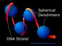
3  Lee, Cameron et. al.  “Designing dendrimers for biological applications” Nature Biotechnology Vol. 23 No. 12 p.1517-1526 (Dec 2005).

4  Supattapone, S. et. al.  “Elimination of prions by branched polyamines and implications for therapeutics.”  Proc. Natl. Acad. Sci Vol. 96 p.14529-14534 (1999).

5  Bourne, N. et. al.  “Dendrimers, a new class of candidate topical microbicides with activity against herpes simplex virus infection”  Antimicrob. Agents Chemother. Vol. 44 p.2471-2474 (2000).

6  Brinas, R.P. et. al.  “Phosphorescent oxygen sensor with dendritic protection and two-photon absorbing antenna ” J. Amer. Chem Soc. Vol. 127 p.11851-11862 (2005).

7  Dunphy et.al. “Oxyphor R2 and G2: phosphors for measuring oxygen by oxygen-dependent quenching of phosphorescence.” Anal Biochem Vol. 310 p. 191-198 (2002).

8  Choyke, P.L. and Kobayashi, H.  “Functional magnetic resonance imaging of the kidney using macromolecular contrast agents.”  Abdominal Imaging Vol. 31 No.2 p.224-231 (April 2006).

9  Wathier et. al. “Dendritic macromers as in situ polymerizing biomaterials for securing cataract incisions” J Amer Chem. Soc  Vol. 126 p.12744-12745 (2004).

10  Sontjens et. al.  “Biodendrimer-based hydrogel scaffolds for cartilage tissue repair”  Biomacromolecules Vol. 7, No. 1 p.310-316 (Jan 2006).