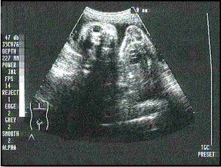
A traditional ultrasound image

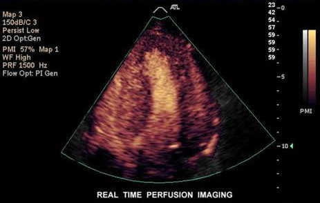
Contrast enhanced ultrasound image
What is traditional ultrasound?
Ultrasound or ultrasonography is a medical imaging technique that uses high frequency sound waves and
their echoes. It is used to form an image of organs or tissue inside the body non-invasively.
How does it work?
The ultrasound machine transmits high-frequency (1 to 5 MHz) sound pulses into the body using a probe.
The probe has special crystals that generate the sound waves and also detect sound waves reflected.
When the sound waves hit a boundary between tissues (e.g. between fluid and soft tissue, soft tissue
and bone) some of the sound waves are reflected back to the probe and the rest travel on further until
they reach another boundary and are reflected back. The reflected waves are detected by the probe
and relayed to the machine. The machine calculates the distance from the probe to the tissue
or organ (boundaries) using the speed of sound in tissue (1,540 m/s) and the time of each echo's return
(usually in the order of millionths of a second). The machine displays the distances and intensities of
the echoes on the screen and forms a two dimensional image.
What is Contrast Enhanced ultrasound?
Contrast enhanced ultrasound (CEU) is the application of ultrasound contrast agents to traditional medical
sonography. Ultrasound contrast agents are gas-filled microbubbles that are administered intravenously
to the systemic circulation.
How does it work?
Microbubbles have a high degree of echogenicity, which is the ability of an object to reflect the
ultrasound waves. The echogenicity difference between the gas in the microbubbles and the soft
tissue surroundings of the body is immense. Thus, ultrasonic imaging using microbubble contrast
agents enhances the ultrasound backscatter, or reflection of the ultrasound waves, to produce a unique
sonogram with increased contrast due to the high echogenicity difference.