Causes of Hearing Disorder
Gene expression
Over 100 genes are thought to be involved in this pathway since the chromosomal locations for almost separate 100 loci have been determined (Hereditary Hearing Loss homepage) and the genes for over 40 have been cloned (Deafness Gene Mutation Database). Some of these genes, for example, CDH23, KCNQ4, MYO15, OTOF, PRES, and STRC, are expressed primarily in hair cells and play a critical role in hair cell physiology. Dysfunction of other cochlear cell types also results in hair cell degeneration, resulting in hearing loss. For example, mutations of GJB2 [1], which is expressed in the supporting cells of the organ of Corti, in the basal cell region of the stria vascularis, and in type I fibrocytes of the spiral ligament, are the leading cause of nonsyndromic hearing loss [2]. Other genes such as TECTA, OTOA, and COL11A2, which are expressed in the tectorial membrane, also are involved in hearing loss and can lead to the degeneration of spiral ganglion neurons [3].
Panels in figure 1 are the scanning electron microscopy images and confocal microscopy images which show the auditory sensory spithelia of wild type (A-D) and mutaed (E-H for Tlc/+, I-L for Tlc/Tlc) mice at P0. Low magnification images (A, E, and I) show that the characteristic arrangement of thee rows of outer hair cells and a single row of inner hair cells is maintained in Tailchaser mutants. In wild type animals, outer (B) and inner (C) hair cell stereocilla form highly organized bundles with kinocillium present at the center of the apical plane. Tailchaser mutants, however, show highly disorganized and misshaped stereocilia boundles on outer (F, and J), and inner (G and K) hair cells. The position of the kinocilium was highly variable in Tailchaser mutants. Confocal microscopy images show the actin in stereocilia bundles (red) and the kinocilia (green). It is apparent that the organization of hair cell bundles in wild type and mutants are quite different. Since we know the structure of hair cell bundle indeed influences the vibration pattern and the regulation of the mechanosensitive ion channel on the inner hair cells, the disorder of hair cell bundles cause the hearing loss is expectable [4].
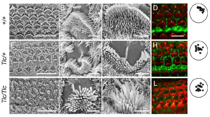
Figure 1 The kinocilia is mislocalized in tailchaser hair cells
Damage of spiral ganglion neurons
Sensorineural deafness is typically caused by irreversible loss of hair cells. Loss of spiral ganglion neurons occurs secondary to inner hair cell loss. Animal studies have shown that spiral ganglion neurons degeneration following hearing cell loss may be either gradual or rapid. Similarly, histological studies of human ears have demonstrated a marked variability between individuals in the extend of spiral ganglion neuron loss following hair cell degeneration. The degeneration of spiral ganglion neurons (SGN) is probably a result of loss of survival factors including the lack of depolarizing activity, which is usually provided by the inner hear cells. [5]
During development, the SGN depend on a variety of neurotrophic factors for survival. Brain-derived neurotrophic factor (BDNF) and neurotrophin –3 (NT-3) have been shown to be critical for normal innervation to the developing inner ear [5]. Additional factors, including glial-derived neurotrophic factor (GDNF) have also been found to improve neuronal survival of developing SGN [6]. Thus, early efforts to restore degenerated, adult SGN have focused on infusing these same growth factors in animal models of sensorineural hearing loss.
Auditory neuropathy (AN) is hearing disorder which is believed to involve damage to the inner hair cells, faulty connections between the inner hair cells and the nerve leading from the inner ear to the brain, or damage to the nerve itself. Figure 2 below shows the comparison of the auditory nerves between two persons with normal hearing function and with AN. There is a marked reduction in the number of auditory nerve fibers in the AN patient. The sural nerve in the patient also showed axonal loss with evidence of demyelination and remyelination. Figure 3 shows the cross sections of sural nerves from normal hearing subject as well as from AN subject. It can be clearly seen that the loss if large myelinated fibers in the subject with AN and the thinning if myelin in many of the remaining large fibers [7].
Figure 2 Photomicrographs of a cross section of the auditory nerve in a patient hereditary sensory-motor neuropathy and another person with normal hearing
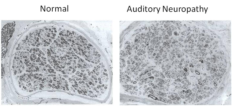
Figure 3 Cross section of a normal sural nerve biopsy and a sural nerve from an auditory neurapathy subject with a concomitant peripheral neuropathy
Other factors
Presbyacusis: it is a kind of age-related hearing loss. This is a natural decline in the hearing. Many people get this as they get older because of damage to the hair cells in the cochlea.
Acoustia trauma: it is the damage to the hair cells by loud noise. This is more likely to happen if people are exposed in a noisy environment for a long while
Infections such as measles, mumps, or meningitis.
Acoustic neuroma: it is a benign tumor affecting the auditory nerve causing deafness and tinnitus
Medicines such as some powerful antibiotics can cause permanent hearing loss. At high doses, aspirin is thought to cause temporary hearing loss and tinnitus
Certain cancer treatment, such as chemotherapy and radiation therapy, can cause hearing loss
However, the loss of hair cells from the human cochlea and vestibular organs is a leading cause of permanent impairment of hearing and balance.
Reference
[1]Denoyelle F, Marlin S, Weil D, et al. "Clinical features of the prevalent form of childhood deafness, DFNB1, due to a connexin-26 gene defect: implications for genetic counselling". Lancet 1999;353:1298–130
[2] Monisha Mukherjee, SR Phadke, Balraj Mittal, "Connexin 26 and autosomal recessive non-syndromic hearing loss", Indian Journal of human genetics, Vol. 9, issue. 2, p 40-50, 2003
[3] Linthicum FH Jr, Anderson W. "Cochlear implantation of totally deaf ears. Histologic evaluation of candidacy". Acta Otolaryngol 1991;111:327–331
[4] Ronna Hertzano, Ella Shalit, Agnieszka K. Rzadzinska, et al. "A Myo6 Mutation Destroys Coordination between the Myosin Heads, Revealing New Functions of Myosin VI in the Stereocilia of Mammalian Inner Ear Hair Cells", PLoS Genetics, Vol. 4, Issue 10, Oct. 2008.
[5] Toshihiko Nakaizumi Kohei Kawamoto Ryosei Minoda Yehoash Raphael, "Adenovirus-Mediated Expression of Brain-Derived Neurotrophic Factor Protects Spiral Ganglion Neurons from Ototoxic Damage", Audiol Neurootol 2004;9:135–143
[6] Ylikoski, J., Pirvola, U., Virkkala, J., Suvanto, P., Liang, X-Q., Magal, E., Altshucler, R.A., Miller, J.M., Saarma, M. "Guinea pig auditory neurons are protected by glial cell line-derived growth factor from degeneration after noise trauma" Hear Res.,1998, 124, 17.
[7] Robert V. Harrison "Auditory neuropathy: Basic science perspectives", 9th Conference Canadian Academy of Audiology Oct 2006 Calgary Canada.