Immunotargeted Nanoshells for Integrated Cancer Imaging and Therapy
Christopher Loo, Amanda Lowery, Naomi Halas, Jennifer West,and Rebekah Drezek
In this study, the researchers used nanoshells to create an integrated imaging and therapeutic breast cancer technology. Nanoshells are essentially silica cores which are covered by a thin shell of metal (in this case, gold). Nanoshell surfaces are quite versatile. They can be “stealth coated” for biocompatibility with polymers such as PEG, or the surfaces can be modified to enable selective targeting of oncogenic proteins that are overexpressed on cell surfaces. In this study, the protein HER2, a protein overexpressed in breast cancer was targeted. The nanoshells also provide a high level of selectivity. Since tumors tend to have leaky vasulature and high retention for small particles, nanoshells tend to accumulate in tumor regions. This, and the anti-HER2 coupled to the nanoshell surface, enhances specificity of binding to tumor cells only.
The optical properties (i.e. scattering and absorption) of nanoshells change when the relative radii of the core and the metal shell are changed. For imaging, a contrast agent or medium must be highly scattering, thereby increasing the fluence rate and giving high resolution. Imaging methods that can be applied with the use of nanoshells are mostly based on photonics, such as OCT. For therapy, an agent must have a high absorption coefficient, so that the energy can be redirected in the form of heat and destroy cancer cells via photothermal ablation. Nanoshells can be made in such a way to have high scattering and absorptive coefficients at certain wavelengths. In this study, the core of the nanoshell was 120 nm and the shell was 10 nm thick. The wavelength was 820 nm near-infrared radiation (NIR), since absorption and scattering efficiencies are highest in this range.
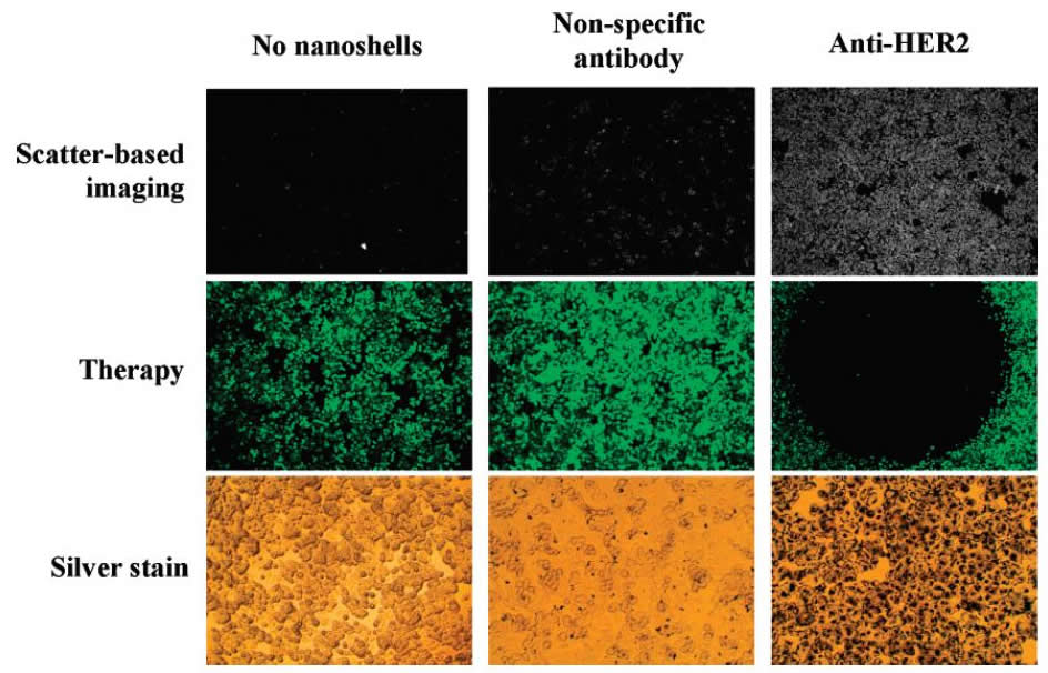
The study sought to simulate the imaging and therapy approach in-vitro. Two controls were used: one with no nanoshells and one with a non-specific antibody added. For the experimental setup, cancer cells that overexpress HER2 were cultured, and nanoshells were added. The 820 nm radiation was then cast incident onto all samples. The samples were first imaged and then ablated. Nanoshell binding and cell viability were stained for by silver stain and calcein (green) respectively.
The results of this experiment were staggering. In the case of the experimental setup, the calcein stain showed cytotoxicity (black) in areas corresponding to high nanoshell binding in the silver stain, whereas the controls showed no such correspondence (see middle-right of figure).
This study is quite encouraging from a diagnostics and therapeutic standpoint. It allows for high resolution imaging using existing photonics imaging technology. The nanoshells can be tuned to different frequencies of wavelengths (since their optical properties can be changed). This allows imaging and ablation to be conducted at NIR wavelengths where absorption and scattering from other chromophores in tissue is minimal. As far as diagnostics goes, the specificity of nanoshell binding is by far the biggest strength. Suspicious masses can very quickly be diagnosed as cancerous if there is a high accumulation of antigen-specific nanoshells that are sequestered in the area. Also, while this may be an expensive method of imaging, the dual nature of this technology allows for cost-cutting in treatment, reducing overall cost.
NOTE: While this is a promising technique, it must be mentioned that HER2/c-erbB-2 is overexpressed in only 20-30% of breast cancer cases. Thus, this technology would have limited application, similar to the ELISA detection case.